Tuesday, November 18, 2008

Acromegaly in MRCP

Acromegaly in MRCP

Acromegaly is always a popular case in MRCP PACES but I think it is an important endocrine illness as well in MRCP Part 1 and 2.

OK, acromegaly is an endocrine disorder with excessive growth hormone, I think everyone knows about that. The name ‘acromegaly’ comes the Greek words for “extremities” and “enlargement,” reflecting one of its most common symptoms—the abnormal growth of the hands and feet. It is easy to diagnose acromegaly in paediatrics patients ( because patients will present with gigantism) however, sometimes you might miss acromegaly in adult group.

Acromegaly is a popular case in MRCP PACES short station. However, for MRCP Part 1 and 2, common questions will be as below,

1) Ways to diagnose acromegaly

Always remember that you need OGTT first and later to confirm with Growth Hormone level. You may need to check insulin-like growth factor 1 (IGF-1). MRI brain is useful as well!

2) Picture test


They like to show you a picture and ask you about the diagnosis. I think no one should fail this because YOU SHOULD NEVER miss acromegaly in your life.


3) Symptoms and signs of acromegaly

Remember that patients may just present with hypertension or diabetes. And of course do not forget about loss of libido as well!

OK, about the treatment – just remember surgery or medical- classes of drug to be used-somatostatin analog and GH receptor antagonist

Tuesday, October 21, 2008

Hypercalcemia in MRCP(2)


Hypercalcemia in MRCP(2)

As a medical student many years ago, I remembered I have to memorize a lot of medical mnemonics. It is easy to remember how a patient with hypercalcemia presents to hospital, just remember this sentence,


“ STONES, BONES, ABDOMINAL MOANS, AND PSYCHIC GROANS”

Let me explain these symptoms briefly,
1) Stone-

I think it is rather straightforward, high calcium in the blood also translates high calcium in the urine, therefore you are prone to get stone. Besides that, patients with hypercalcemia also easily get dehydration because they might have polyuria due to nephrogenic diabetes insipidus.

2) Abdominal moans-

Hypercalcemia leads to constipation, abdominal colic and pancreatitis.

3) Bones-

You get bone pain because there is increased in bone resorption/ breakdown due to tumour ( causing pathological fracture) or hyperparathyroidism.

4) Psychic groans-

I can’t explain this, hypercalcemia can cause psychosis, confusion etc. You have to remember it!!But I think all the electrolyte imbalances can cause some kind of mental problems.

About the treatment of hypercalcemia, I think it is not so important to remember, anyway, remember the following strategies,
1) Rehydration
2) Steroids
3) Calcitonin
4) Biophosphonates
5) Plicamycin
6) Dialysis
7) And of course, treat the underlying cause!!

However, just want to remind all of you, there are a few causes of hypercalcemia that you might can’t explain the mechanism involved but they are important. These causes are thyrotoxicosis, Addison’s disease and acromegaly.

If you can explain the mechanism involved, please share with other readers!!

Wednesday, October 08, 2008

Hypercalcemia in MRCP (1)

Hypercalcemia in MRCP

As a houseofficer many years ago, I remember that there are two electrolytes that are frequently encountered during clinical practice- Potassium and Calcium.

We have discussed a lot about Potassium, I am going to talk about Calcium metabolism today and of course talk more about hypercalcemia.

It is pretty easy to remember, the only pool of calcium in our body is bone. Although tiny amount of calcium is being absorbed through the gut ( affected by Vitamin D), maintenance of normal calcium level in serum ( 2.2-2.6) greatly depends on exchange of Calcium between extracellular fluid and bone.


It is easy to remember that if we have low calcium level, our body will try to do the followings to increase calcium level in the serum,

1) Increase Calcium absorption from the gut
2) Increase bone resorption in the bone so that more calcium can be released to the serum
3) Reduce Calcium excretion from the kidney

The main organ that regulates these is parathyroid hormone. You can think of causes of hypercalcemia into a few big groups as below,

1) Bone problem
It is easy to understand this, when there is increased bone destruction, of course you calcium level is high. Therefore, any malignant disease either primary or secondary that leads to bone destructions can cause hypercalcemia.

2) Vitamin D problem
As I said before, Calcium absorption from the gut is mainly affected by Vitamin D, therefore, Vitamin D toxicity or granulomatous diseases ( such as Sarcoidosis or tuberculosis) can cause hypercalcemia.

3) Parathyroid hormone
Of course, when you have high parathyroid hormone ( primary and secondary), you calcium level is high but remember that secondary hyperparathyroidism may have normal or even low Calcium level.

4) Others
Some other rare causes such as Familial hypocalciuric hypercalcemia, milk alkali syndrome, immobility etc.

Saturday, September 20, 2008

Immunosuppressive Drugs (1)

Immunosuppressive Drugs- Cyclosporin


Sorry for the long absence from my blog. I just shifted to my new house and had to live without broadband for almost 3 months.


OK, today I am going to talk about cyclosporin ( prototype of calcineurin inhibitor) because this drug change the landscape we look at solid organ transplantation. It was discovered in 1971 and subsequently approved for use in 1983.


I do not think you care about the history. The more important topics you want to know are popular questions in MRCP, here are the popular questions,


1) Drug Interacations

Since cyclosporin is metabolised in the liver by cytochrome P-450, there are a lot of drug that can induce/inhibit this enzyme causing low/high cyclosporing level in the blood. Fo the mneumonics of enzyme inducers/inhibitors, you can read my previous blog. Remember that grape juice inhibit the cytochrome P-450!! ( ALL-TIME POPULAR MRCP QUESTION!!)


2) Side effects


This topic is ver popular if you get a case of kidney transplant in MRCP PACES, common side effects are,

Tremor
Hypertension
Gum hypertrophy
Electrolyte imbalance
Nephrotoxicity

I will talk more about immunosuppressive drugs in my future blogs!!

Monday, July 21, 2008

Guillain Barre syndrome in MRCP

Guillain Barre syndrome in MRCP


There are only a few common neurology problems that are popular in MRCP. One of them is Guillain Barre syndrome and I think I will try to highlight some salient points about this condition.


First thing to remember about this condition is we always term any medical problem a syndrome when we do not understand fully about it.


GBS was first described in 1859 by Landry. Guillain Barre syndrome is a type of acute inflammatory demyelinating peripheral neuropathy mainly involving the motor modality.


Although it may involve sensory or autonomic modality, classically you will be given a question involving motor neuropathy in MRCP.


GBS is believed to result from autoimmune humoral- and cell-mediated responses to a recent infection or any of a long list of medical problems.


Second lesson to be learned if you are sitting for MRCP is patient with GBS usually come to the hospital after viral or bacterial infection. The common infections associated with GBS are Campylobacter jejuni , Haemophilus influenzae, Mycoplasma pneumoniae, and Borrelia burgdorferi and influenza. Therefore patients usually have gastrointestinal and respiratory illness before the onset of GBS.

Patient with unilateral foot drop

Patients usually come with ascending weakness and some of them may complain numbness over the extremities.The classical physical signs are bilateral foot drop with loss of reflexes. However, remember some rare variants involving cranial nerves may be seen ( Miller-Fisher),patients may present with facial weakness mimicking Bell palsy, dysphagia, dysarthria, ophthalmoplegia, and pupillary disturbances.


Patients with GBS will usually die because of autonomic dysfunction with cardiac dysrhythmias or respiratory muscle involvement.
How to diagnose GBS, you have to do lumbar puncture, classically you will find elevated CSF protein. However, you may want to do nerve conduction study ( a delay in F wave), if you are suspecting Miller-Fisher, anti-GQ1b may be present.
How to monitor your patient’s respiratory function, monitor their Forced vital capacity.


Treatment is giving IV Immunoglubulin!

Monday, May 05, 2008

Liver Cirrhosis in MRCP

Liver Cirrhosis in MRCP


I came across a lot of liver cirrhosis cases during my housemanship. I remember a patient who actually came to medical ward almost every month for theraupeutic peritoneal tapping.
Liver cirrhosis just means the liver is irreversibly destroyed by fibrosis and degeneration of the hepatocytes. Actually, it should be a pathology diagnosis, however we always can diagnose this by physical signs and ultrasound alone.
There are a few important points for you to remember if you are sitting for your MRCP Part 1 and 2, I would summarize these points as below,


1) Causes of liver cirrhosis
The causes of liver cirrhosis greatly depend on where you are working. If you work in Western countries, alcohol is always the number one cause. However, chronic hepatitis will be top in the list if you live in Asia. For your MRCP, there are three more causes you need to remember- cryptogenic ( idiopathic), Budd-Chiari syndrome and haemochromatosis. I talked about haemochromatosis before, please read about it.


2) Clinical signs of chronic liver disease
If you are studying for your MRCP PACES, then you will know that there are more than 20 signs for stigmata of chronic liver disease. However, remember a few important ones such as jaundice, spider naevi, gynaecomastia, testicular atrophy, leuconychia, finger clubbing.......etc

3) Investigations
First you must try to find out the underlying cause, second you must prognosticate your patient. Child’s criteria is the important criteria to remember. The mnemonic to remember- BAPA + E( BAPA means ‘father’ in Malay language)- Bilirubin level, Ascites, PT ( INR) and Albumin level and encephalopathy.

4) Complications of liver cirrhosis
Patients usually die because of upper GIT bleeding. However, they are bought to hospital because of hepatic encephalopathy. Remember all the precipitating of hepatic encephalopathy.

5) Treatment of liver cirrhosis
Almost all are supportive, liver transplantation provides cure but almost not done in this part of the World. However, always prevent hepatic encephalopathy and minimize the risk of UGIB. Do yearly monitoring to look for liver cancer.

Monday, April 28, 2008

Gyanecomastia for MRCP

Causes gyanecomastia for MRCP

I was asked by a medical student about gyanecomastia today during my ward round.
I think it is important during your MRCP Part 1 because it is a popular question. OK, before talking about the causes, let us define what gyanecomastia is. Gyanecomastia just means male breast enlargement.

It is certainly abnormal for male to get breast enlargement, however, you may be suprised that I divide gyanecomastia into physiological and pathological gyanecomastia.

You heard me right, there are times in a male life that he can get breast enlargement abd it is totally physiological!

Man gets gyanecomastia when they are newborn, adolescents (puberty) and they are old!
Causes of pathological gyanecomastia are enormous, however there are only a few big groups,

1) Drug related
I always remember a few important ones, they are cimetidine, ranitidine ( H2 antagonists), spirolactone, digoxin and of course estrogen or drug that makes you less masculine.

2) Certain tumours
Popular ones are brochogenic carcinoma, testicular tumour and HCG producing tumours

3) Congenital
Popular syndromes are Klinefelter syndrome, Kallman syndrome

4) Systemic illness
Popular systemic illnesses are chronic liver disease and in certain chronic kidney disease.

Thursday, April 10, 2008

Chronic Myeloid Leukemia in MRCP

Chronic Myeloid Leukemia in MRCP

Chronic Myeloid Leukemia (CML) is always a popular differential diagnosis in your MRCP PACES examination if you encounter massive hepatosplenomegaly during your abdominal short case.

CML is one of the 4 disorders ( besides polycythamia rubra vera, essential thmrobocythemia and myelofibrosis) termed as myeloproliferative disorders.

The term myeloproliferative disorders describes a group of conditions characterized by clonal proliferation of one or more haemopoietic components in the bone marrow and in many cases, the liver and spleen.

OK, patients usually present the following ways,

1) abdominal pain and distention because of massive hepatoslpenomegaly
2) bleeding tendency due to platelet dysfunction
3) features of anaemia
4) gout or renal impairment due to hyperuricaemia ( because of excessive purine breakdown)
5) some rare symptoms such as priapism ( this is the only cause of priapism I can remember during my medical school time because there was no Viagra yet at that time!!)

I think if you see a case of CML during your MRCP PACES, you must know how to come to a diagnosis of CML, basically, you can do the following,

1) You always find very high total white cell count when you do full blood count. I remember when I was a house-officer, I encountered a patient who were well and had a TWC of 150,000!!
2) Neutrophil alkaline phosphatase ( NAP) score is low!! ( Remember this well because it is a popular question in MRCP. Also remember diseases that have low NAP score!)
3) Chromosomal study- Remember that you usually find Philadelphia Chromosome which is a translocation of chromosome 9 and 22. ( This is the hottest exam question in MRCP and also your final MBBS!!)
4) Bone marrow is hypercellular with granulopoitic predominance.
5) Peripheral blood film may show various stages of granuloiesis including promyelocytes, myleocytes, metamyelocytes and band and segmented neutrophils


When I was a house officer, I remember that my consultant used a lot of hydroxyurea to treat CML. However, currently imatinib ( Gleevec) which is a tyrosine kinase inhibitor has become the first line treatment for CML.

Saturday, March 22, 2008

Hyperkalemia in MRCP (2)

Hyperkalemia in MRCP (2)

OK, if you are currently working in any hospital around the world, you certainly agree with me that hyperkalemia always disturbs you a lot. I remember that when I was a house officer many years ago, I was once called by staff nurse because she was worried that patient may collapse simply because his Potassium level was 5.4!


I think the general principles of treating hyperkalemia are simple,

First,

You have to act fast to avoid cardiac arrhythmia.

Second,

To shift the Potassium back to the cell ( intracellular) from extracellular ( plasma) if possible.

Third,

To reduce total body Potassium

Fourth,

AND of course, find out the underlying cause of hyperkalemia.


I will not discuss how aggressive you want to treat hyperkalemia and I think it is a judgment call. Anyway, I would be certainly very worried if the Potassium level is more than 6.5 and there is ECG changes. ( Learn about hyperkalemia associated ECG changes, it is a popular question in MRCP).

So, treatment of hyperkalemia can be outlined as below,

I think the very first step to take is to stabilize the heart by giving Calcium gluconate or Calcium chloride. You may want to open your physiology book to learn the mechanism how Calcium acts.

Then ,of course, you want to try to shift back the Potassium back to the cell by giving insulin and glucose. In Malaysia, the combination of insulin, glucose and Calcium therapy in treating hyperkalemia is termed as cocktail regime!

You can also use beta agonist to shift the Potassium back to the cell. Another useful strategy is bicarbonate infusion. Remember, acidosis causes hyperkalemia, therefore alkalosis corrects hyperkaelmia.
Another strategy you may want to try is giving patient cation exchange resin. However, remember that the effect is not immediate, therefore, you have to use previous various strategies to bring down the Potassium level promptly.

Anyway, I must say the most powerful way of treating your hyperkalemia is haemodialysis!!

Thursday, March 13, 2008

Hyperkalemia in MRCP (1)

Hyperkalemia in MRCP – Part 1

Electrolyte imbalance is an important topic in MRCP and I think Potassium is the single most important electrolyte in our bodies.

If you are a house officer, I think the commonest electrolyte abnormality you will see in medical ward is hypo/hyperkalemia.

Today, we will discuss about hypokalemia. Before we talk further about causes of hyperkalemia and how do we manage this, we have to learn about basic physiology.


First fact to remember, potassium is mainly intracellular, the concentration of K is about 150mmol/L of H2O inside the cell as compared to about 5 mmol/L outside the cell ( in plasma). Therefore, to maintain this concentration, our body depends greatly on Na-K ATPase channel ( this channel transport 3 Na out of the cell for each 2 K it transports in), however , you must always remember there are H-K ATPASE in specific organs such as kidneys for similar purpose.

Potassium is mainly excreted in kidney although a small proportion is excreted through GIT.

OK, let us talk about causes of hyperkalemia, I can divide them into either increased load, reduced excretion and increased release from cells ( Remember? Potassium is mainly intracellular!)

1) Reduced excretion
Chronic kidney disease ( Potassium is mainly excreted via kidney )
Mineralcorticoid deficiency ( learn the effect of mineralcortiocid on Na-K channel, you will understand)
Some drugs ( especially ACEI/ARB, heparin, potassium sparing drug)

2) Increased load
Overzealous Potassium supplement
Transfusion of blood

3) Increased release from cell
Any causes leading to major cell breakdown such as tumour lysis sundrome, tissue necrosis, rhabdomyolysis
Acidosis ( Remember I told you about H-K pump!!)
Beta blocker

OK, I will talk about management of hyperkalemia in my next post.

Saturday, February 16, 2008

Hyperthyroidism in MRCP

Hyperthyroidism in MRCP

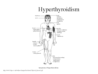
Hyperthyroidism is the commonest endocrine problem you will see during your practice either you are in endocrine unit or general medicine.

Therefore, I think you must learn hyperthyroidism well and it is commonly asked in your MRCP/USMLE examination as well.

Common causes of hyperthyroidism are Grave’s disease, toxic multinodular goiter and toxic nodule (adenoma). You will most probably seeing mostly Grave’s disease as the cause of your patient’s hyperthyroidism especially among younger female patients.

Anyway, first thing to remember in your MRCP, there are a lot of drugs that can cause hyperthyroidism and two commonly asked drugs are Lithium and amiodarone. I have talked about amiodarone in my previous post. Learn this drug hard because it is important and a popular drug in your exam.

OK, to learn about the signs and symptoms of hyperthyroidism, it is rather logicaland easy to remember. It is an important metabolism hormone, therefore when there is an increased level of thyroid hormone, everything in your body is increased- your heart rate, your metabolism rate, your gut peristalsis etc. Therefore, you anticipate patient to compliant palpitation, weight loss and diarrhoe. Depending on whether patient has Grave’s disease, you may get some eye symptoms and signs.


However, as a medical student before, I remember that everything in hyperthyroidism is increased except patients have reduced power ( proximal myopathy) and female patients may have less/reduced menses ( amenorrhoea). Remember as well urticaria can develop in hyperthyroidism.

Another thing to remember, a lot of young patients with hyperthyroidism have a lot of symptoms but always older patients with hyperthyroidism appear to be ‘silent’ ( no symptoms) and they always present with just atrial fibrillation or symptoms suggesting heart failure.

One lesson to be learned here, always check patient’s thyroid function if you can’t find out the underlying cause of patient’s heart failure especially among older patients.

Learn how to interpret thyroid function test ( easy, T3 or T4 is high with low TSH suggest hyperthyroidism), however, remember the side effects of anti-thyroid drugs.
Two important side effects to remember- agranulocytosis and skin rash. It is your duty to check patient full blood count after you start them on anti-thyroid drugs because patient may later come to you’re A+E with leucopenic sepsis due to carbimazole!

Wednesday, January 23, 2008

Prolactin in MRCP

Prolactin in MRCP

I always enjoyed studying endocrinology during my medical school time. One of my old professors said, endocrinology is straight forward and logical. Our body is designed in a way when our hormone level is high, there will be a negative feedback and vice versa. Our bodies try to maintain a normal level of all hormones so that we can function normally.

A trick to remember when you study endocrinology, you must understand normal physiology so that you can understand each hormone clearly and not just memorize them by hard.

OK, today, we will start to learn the first hormone- prolactin. Why prolactin?It is rather interesting that we know prolactin is important for females because it helps in milk production but its function in males remains a mystery!

I think there are a few important facts about prolactin that always asked in your MRCP!

Fact 1:

All hormones in pituitary glands are up regulated by another hormone in hypothalamus ( positive feedback) except prolactin. This means that prolactin production will be inhibited by another hormone prolactin inhibiting hormone ( PIH) from hypothalamus. Remember that PIH is dopamine, therefore your dopamine agonist such as bromocriptine is used to suppress prolactin and thus milk production. ( And also remember that due to its dopaminergic effects, bromocriptine is used in Parkinson’s disease).

Whereas drugs which as anti-dopamin effect such as metoclopramide is used to stimulate prolactin production and it is always used in O+G for post partum mothers if they have problems in milk production.

Fact 2:

You do not believe it, prolactin is a stress hormone. As a medical student, I always do not understand why God created prolactin as stress hormone. Anyway, prolactin level can be measured if you want to differentiate a true seizure from pseudo-seizure because its level is high after an epileptic fit.

Fact 3:

When there is a non-secreting tumour in pituitary causing damage to the stalk, you anticipate secretion of all hormones from pituitary to be reduced ( because positive feedback from hypothalamus) but prolactin level is high because there is no negative feedback from hypothalamus.

Fact 4:

One of the commonest causes of hyperprolactinoma and galactorrhoea is drug-induced and it is due to Phenothiazines!!

Friday, January 11, 2008

Benign Intracranial Hypertension in MRCP

Benign Intracranial Hypertension in MRCP

I always remember that benign intracranial hypertension is a popular topic in MRCP Part 1 and 2. Recently, my wife was studying her FRACGP and I noticed that BIH is one the hottest topics as well.

Since this illness is so popular and important, I think we should spend sometime talking about BIH today.

OK, why we say intracranial hypertension is benign?? When there is intracranial hypertension, we anticipate there will be some problems inside our craniums, however, if there is presence of intracranial hypertension without any obvious intracranial mass or enlargement of ventricles or hydrocephalus, we term the illness as BENIGN ( it won’t kill you!!) intracranial hypertension.

There are a few facts to remember for BIH,

Fact 1:


Remember that majority of patients are young female who are obese and usually in your MRCP, they will give you an example of an obese lady with acne. Why acne?? I always wondering when I was a medical student. After struggling for many years, I finally understood this. The reasons are, some anti- acne actually cause BIH such as teteracycline, Vitamin A and drugs that can precipitate acne formation such as steroid also lead to BIH!!

Fact 2:

Although we were taught that papilloedema is an emergency if patient has headache. Remember that patient with BIH has headache and papilloedema ( although rarely they might have blurring of vision and seizure) but it is benign and the brain imaging and CSF are normal.

Fact 3:

Since patient with BIH is always a young female patient, you must put sagittal sinus thrombosis as your differential diagnosis. This is because you also anticipate young ladies are prone to get autoimmune disease especially SLE and they are usually on oral contraceptive pills and these put the ladies at risk of developing sagittal sinus thrombosis.


Fact 4:

Treatment is easy, stop the drug and weight reduction but you may use loop diuretics or acetazolamide.


Example of question:


A 22-year-old obese woman presented with an 8-week history of headaches, pulsatile tinnitus and transient visual loss on standing lasting a few seconds. She had otherwise been well with no history of note. She took the oral contraceptive pill and had been taking this for the last 6 months and used salbutamol inhalers on an occasional basis for her asthma which she had from childhood. She also took vitamin Asupplements which she bought over the counter for her general health. On examination, the only abnormality of note was bilateral papilloedema. MRI brain and MR Venogram are normal. Lumbar puncture showed an opening pressure of 38, normal protein, glucose, and cells.. What is the most likely diagnosis?


1 )Herpes simplex encephalitis

2 )Intracranial hypertension secondary to vitamin A

3 )Malignant meningitis

4 )Sagittal sinus thrombosis secondary to OCP

5 )Sagittal sinus thrombosis secondary to SLE

Tuesday, January 08, 2008


Drug in MRCP-Phenytoin

Although some of you may be not so familiar about phenytoin especially for those who are practicing medicine in developed countries. I think this is because there are so many new antiepileptic drugs available in the market now.

Actually, phenytoin is the oldest non-sedative antiepileptic drug introduced in 1938!!
I think it is not so important for you to understand how phenytoin acts because I myself never understand it when I was a medical student myself many years ago.

In MRCP examination, there are a few important facts that you must always remember.

Fact 1 : Drug metabolism/binding

Remember that phenytoin is mainly bound to protein. Therefore, when there is hypoalbuminemia, there is decreased protein binding- results in a decrease in total plasma concentration of drug but not the free concentration.

Therefore a lot of doctors tend to increase the drug dosage to maintain total drug levels in the therapeutic range- leading to toxicity.

Besides that remember that hepatic enzyme induction and inhibition also alter its drug level.

Although phenytoin is mainly metabolized in liver, its metabolites are excreted in kidney, therefore, renal failure may precipitate toxicity.

Fact 2: Side effects

As I remember as a medical student, there are two interesting side effects of phenytoin- gum hypertrophy ( Look out the photo at http://www.passpaces.com/ ) and generalized lymphadenopathy. However, remember that acute toxicity of phenytoin also leads to cerebellar signs!!

Fact 3: Cardiac complications

Since phenytoin alters Na, K and calcium conductance, it can cause cardiac arrhythmia, therefore always put patient on cardiac monitor if you suspect toxicity.

Also remember that chronic use of phenytoin can lead to Vitamin D metabolism abnormalities and osteomalacia.