Tuesday, October 31, 2006

Cardiac Catheterization in MRCP(3)

Cardiac Catheterization in MRCP (3)

Today I am going to talk about the last part of this important topic- Cardiac Catheterization in MRCP. In my previous two posts, we have learned about some common cases concerning cardiac catherization in MRCP. I am going to show two more cases here,

Case 1:

Coarctation of the aorta


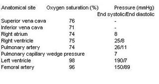
This is an easy case!There is a steep systolic gradient between the left ventricle and the femoral artery; the gradient is calculated as 190 – 150 = 40 mmHg. Therefore the diagnosis is Coarctation of the aorta.

Case 2:

Tetralogy of Fallot


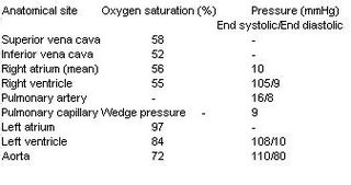
You notice a combination of pressure and oxygen saturation abnormalities. The abnormalities are,

i) Step-down in oxygen saturation between LA and LV, indicating right to left shunt at the level of the ventricles, therefore there is presence of VSD.
ii) Pulmonary stenosis: there is an 89 mmHg gradient across the pulmonary valve (RV systolic – PA systolic).
iii) RVH: Right ventricular pressures are high and there is a right to left shunt, as indicated by the oxygen saturations

Hope you find this information useful. Remember, in MRCP examination, you may be given the result of a cardiac catheterization and asked about the correct physical signs of a patient. In other type of question setting, you maybe given some important physical signs and the examiners want you to find the correct cardiac catheterization results.

Monday, October 23, 2006

Poisoning in MRCP(III)

Poisoning in MRCP (III)-Tricyclic Antidepressants

The third serial of this topic- poisoning in MRCP, I am going to talk about tricyclic antidepressants. Examples of tricyclic antidepressants are amitriptyline, imipramine, doxepin etc.It is a common case scenario you would see during your internship/ housemanship. The reason is simple, doctors prescribe tricyclic antidepressants for patients with depression and these patients tend to use drugs for suicide.

As you might remember, tricyclic antidepressants help depressed patients through inhibition of reuptake of noradrenaline or serotonin in the brain, however, there are a few other effects of tricyclic antidepressants that explain its side effects,


1) it has central and peripheral anticholinergic effects
2) it causes depression of cardiac contractility
3) it slows down intraventricular and artrioventricular conduction ( cardiac conduction)
4) it causes CNS toxicity such as agitation, confusion , coma and seizures


Symptoms of Toxicity

Remember that all these symptoms are due to cardiac toxicity and anticholinergic effects of tricyclic antidepressants.

Cardiac toxicity
Cardiac arrhythmias, supraventricular or ventricular arrhythmias leading to hypotension, pulmonary oedema, therefore patients may present palpitation and breathlessness

Anticholinergic toxicity ( autonomic toxicity)
Dry mouth, urinary retention, dilated pupils, constipation and hyperreflexia

CNS toxicity
Seizures ( may cause severe metabolic acidosis), coma, confusion

Treatment

Gastric lavage
Continuous cardiac monitoring is mandatory
IV Sodium Bicarbonate may be useful to maintain arterial PH if patients develop severe metabolic acidosis
DC shock may be needed, however, anti-arrhythmias are contraindicated
Fluid resuscitation if hypotension
Haemodialysis is not useful.

Tips for MRCP
1) Remember that if you see a patient with dilated pupils, confusion and cardiac arrhythmias , always consider tricyclic antidepressants.

Saturday, October 21, 2006

Rheumatology Questions in MRCP

MRCP Rheumatology Questions

Let's us take a break and look at some of common rheumatology questions in MRCP,

1) A 30 year gentleman was admitted to ward due to history of backpain for 3 months. He denied small joints pain and history of family members having the same problem. There was no history of dysuria and red eye.Below is his X-ray. What is the diagnosis?


1) Ankylosing Spondylitis
2) Riter’s syndrome
3) Psoriatic arthropathy
4) Enteropathic spondylitis
5) Seronegative Rheumatoid arthritis

2) This gentleman presents with swollen joint and fever.


What is likely to be found on microscopy of aspirated synovial fluid?

1 )Bipyramidal crystals that exhibit strong positive birefringence under polarised light
2 )Gram positive cocci in clusters
3 )Needle-shaped crystals that exhibit strong negative birefringence under polarised light
4 )Rhomboid crystals that exhibit weak positive birefringence under polarised light
5 )Small, non-birefringent crystals visible only under electron microscopy

3) A 30 year-old man is admitted to casualty with a 24 hour history of a painful and swollen right knee. He denies any previous history of joint problems. Over the last two days, he has also noticed redness and soreness in both eyes. He has returned from a business trip to Kuala Lumpur a fortnight ago.
On examination, his temperature is 38.5°C. His eyes are red. His right knee is hot, swollen and tender to palpate. No other joint appears to be affected.
Investigations:
Hb 12.9 g/dl
WBC 14.0 x 10⁹/l
Platelets 200 x 109/l
ESR 75 mm/h

Blood cultures
No growth after 48 hours

Urinalysis
No blood, glucose or protein detected

Knee x-ray
Soft tissue swelling around left knee
What is the most likely diagnosis?

1 )Gout
2 )Gonococcal arthritis
3 )Reiter's syndrome
4 )Rheumatoid arthritis
5 )Viral arthritis

4) Which of the following statements are correct regarding this patient's condition?


1 )It occurs more commonly in men
2 )Rheumatoid factor is positive in >90% of cases
3 )It is associated with an erosive arthritis
4 )Raynaud's phenomenon is a feature in ~10%
5 )It is associated with a reduced transfer factor

5) A 22 year old lady presents with typical erythema nodosum. She has a low grade fever and bilateral ankle arthritis but no other symptoms and has no medical history. There is no history of travel abroad and she is on no medication. Which of the following would be the most appropriate investigation for this patient?

1 )Barium enema
2 )Chest x-ray
3 )ESR
4 )Upper GI endoscopy
5 )Viral titres

Get your answers here!

Tuesday, October 10, 2006

Cardiac Catheterization in MRCP(2)

Cardiac Catheterization in MRCP (2)

In my previous post, I talked about a few important examples of cardiac lesion that give you abnormal oxygen saturation, today I am going to give you a few important cardiac lesions ( mainly valvular lesions ) that will give you abnormal pressure during cardiac catheterization.

Before we proceed , remember the normal cardiac pressure and oxygen saturation


Case 1: Mitral Stenosis

Look at the following cardiac catheterization result of a 40-year old lady

The diagnosis is mitral stenosis, you notice a few abnormal results here,

i) The catheter data show a gradient across the mitral valve (LA pressure – LV end diastolic pressure) .Remember that usually LA pressure equals to LV end systolic pressure; you can use the PCWP as a surrogate for LA pressure. In this case the gradient is 26-6 = 20 mmHg.
ii) There is also evidence of right ventricular hypertrophy, with markedly elevated RV pressures due to secondary pulmonary hypertension.

Case 2: Aortic Stenosis

A 65- year old lady was admitted to hospital due to syncopal attack, below is the cardiac catheterization results.

There is a systolic gradient of 81 mmHg across the aortic valve (LV systolic pressure – aortic systolic pressure), indicating severe aortic stenosis. Remember that hypertrophic cardiomyopathy also presents with a similar result, howeve, patients tend to be younger!

Case 3: Aortic regurgitation

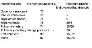
This is an easy case, you notice a wide pulse pressure ( aorta :150/40), therefore the diagnosis is aortic regurgitation.

I would talk about a few more cases in my last post.

Saturday, October 07, 2006

AIDS Defining Conditions in MRCP

AIDS Defining Conditions in MRCP-Kaposi's Sarcoma

I talked about AIDS/HIV three months ago in my blog. Today, we are going to revisit this common disease again because it is a very popular disease asked in MRCP.

There are a few simple facts to remember about HIV,

1) It is a retrovirus ( Family: Retroviridae) . HIV is completely dependent upon CD4 cells for replication and survival.
2) HIV leads to a progression fall in T-helper cells ( CD4) and a failure of T-cell proliferation.
3) HIV infection can be divided into 3 stages- acute seroconversion ( patients usually present with viral-like fever) , intermediate stage ( asymptomatic) and advanced stage ( AIDS)

Advanced stage of HIV (AIDS) is defined when the patient’s CD4 counts drop below 200 /cmm.

When our CD4 drops below 200, we are prone to get all kinds of rare infections ( opportunistic infections) which are uncommon in immune competent hosts.

There are a few AIDS defining conditions that are commonly asked in MRCP examination, these conditions are,

1) PCP
2) Toxoplasmosis
3) Cytomegalovirus
4) Kaposi’s Sarcoma
5) Cryptococcus meningitis
6) Extrapulmonary tuberculosis and atypical mycobacterium
7) Non-Hodgkin’s lymphoma


and others, however, I think these are the most important diseases you must learn.

About PCP, I think I have covered adequately in my previous post. Today I am going to talk about Kaposi’s Sarcoma (KS),

OK, this disease was considered to be very rare before the era of AIDS. It mainly affects elderly men of Mediterranean or Jewish heritage, organ transplant patients, or young adult African men. KS was named for Dr Moritz Kaposi who first described it in 1872.

In patients with AIDS, this tumuor tends to develop in the tissues below the skin surface, or in the mucous membranes of the mouth, nose, or anus. It is always described as raised blotches or lumps that may be purple, brown, or red. Sometimes the disease causes painful swelling, especially in the legs, groin area, or skin around the eyes. KS is caused by a herpes virus called Human Herpes Virus 8 (HHV-8).

However, KS is rare among Asian patients with AIDS as compared to Western patients.
In the skin, KS may not have to be treated if there are only a few lesions. Skin lesions can be:

Frozen with liquid nitrogen,
Treated with radiation,
Cut out surgically,
Injected with anti-cancer drugs or interferon alpha


A few photos of KS are shown below,


Tips for MRCP,
1) Remember that you may not only see KS occurs at skin but also mucous membrane

Tuesday, October 03, 2006

Spondyloarthropathies in MRCP (1)

Spondyloarthropathies in MRCP (1)

This is a group of disorders with following characteristics, ( remember that spondyloarthropathies are popular questions in MRCP.

1) they are seronegative ( Rheumatoid factor negative)
2) usually larger joints are involved such as knees, ankles and sacro-iliac joints
3) if peripheral joints are involved, they are usually asymmetrical
4) characteristic articular features include enthesitis ( inflammation at sites of tendon insertion),dactylitis and scaroilitis.
5) strong association with HLA-B27

The arthropathies that are under this group are,
1) Ankylosing spondylitis
2) Reiter’s syndrome
3) Psoriatic arthropathy
4) Inflammatory Bowel athropathy ( Enteropathic arthritis)

Today I am going to talk about Ankylosing Spondylitis, it is a common short case as well if you are sitting for MRCP PACES, find more discussion at PassPACES.com

Clinical Presentations

Back pain and stiffness, usually happens during the third decade
Peripheral joint pain ( less common )
Uveitis

Physical Signs

Reduced spine movement and chest expansion
‘Question mark’ posture
Anterior uveitis
Anemia of chronic disease
Aortic regurgitation murmur
Achilles tendinitis
Apical fibrosis

Investigations

Sacro-iliac joints involvement


Spine xray- loss of lumbar lordosis, Bamboo spine ( calcification in anterior and posterior spinal ligaments)
Enthesitis
HLA-B27 positive ( about 90%)

Treatment

NSAID, NSAID, NSAID + physiotherapy
Disease modifying drugs have no effect on central disease ( spine) and maybe useful in peripheral disease ( peripheral joints involvement!)

Tips for MRCP,

1) They may show a spine x-ray or pelvic xray with classical history of ankylosing spondylitis, remember how to look for Bamboo spine and sacro-iliac joint involvement!