Tuesday, December 26, 2006

ECG in MRCP(1)

ECG in MRCP(1)

Today I am going to give you a few examples of abnormal ECGs that are commonly asked in MRCP. However, remember a few basic facts first. Look at a normal ECG first!


1) Heart axis- if I and II are both ‘positive’, axis is normal.
2) If every P wave is followed by ORS, then it is sinus rhythm.
3) Normal PR interval is 0.12-0.20 sec.
4) Normal ORS width is less than 0.12s, if it is more than more than 0.12s, it suggests conduction defects.
5) QT interval- From QRS start to end of T wave. Usually QTc is used and it equals to QT interval divided by square root of RR interval. Normal value:0.35-0.43. ( I try to remember as 0.33-0.43, it is a popular question in MRCP Part 1 about causes of prolonged QT interval!)
6) ST segment: either elevation or depression implies infarction or ishaemia.

OK, today I am going to talk about abnormal heart axis. There are two types of abnormal axis either left axis deviation or right axis deviation.

1) Right axis deviation- You will know it if Lead I is negative.


Causes: All pathology causing right ventricular hypertrophy or strain such as cor pulmonale, pulmonary embolism.

2) Left axis deviation- You will notice this if Lead II and III are negative.


Causes: Left ventricular hypertrophy or strain. Such as hypertension, aortic stenosis.

Thursday, December 21, 2006

Merry Christmas and Happy New Year

Merry Christmas and Happy New Year


To all readers of this blog, I would like to express my heartfelt gratitude to all of you for being so supportive to this blog. I promise I will try to make this blog better and provide more useful MRCP tips to all of you!

" Merry Christmas " and " Happy New Year"

Make a wish now and hope you can pass your MRCP in 2007!!

Friday, December 01, 2006

Hyperparathyroidism in MRCP

Hyperparathyroidism in MRCP

OK, today we are going to discuss an important topic in endocrinology in MRCP, we are going to talk about hyperparathyroidism.

Hyperparathyroidism just means high levels of circulating parathyroid hormone. You must remember that there are three types of hyperparathyroidism, primary, secondary and tertiary.

1) Primary hyperparathyroidism
It is simple, there is no reason ( such as low Calcium level) for the raised parathyroid hormone. It is usually due to problems occurring to the parathyroid gland itself. Causes are adenoma, parathyroid hyperplasia or carcinoma.

2) Secondary hyperparathyroidism
Parathyroid gland is stimulated to secrete more parathyroid hormone due to some underlying reason such as low calcium because of renal failure, Vitamin D deficiency.

3) Tertiary hyperparathyroidism
After prolonged stimulation, the parathyroid gland becomes autonomous and secrets parathyroid hormone without any feedback mechanism

I hope to discuss more about primary hyperparathyroidism today because clinically, we see secondary and tertiary hyperparathyroidism mainly in renal failure patients.Therefore, they are not so important in your MRCP Part 1 and 2!

Presentation of Hyperparathyroidism

As you might still remember as a medical student, parathyroid hormone's main function is to mobilize calcium from bone ( therefore leading to hypercalcemia) and increase urinary phosphate excretion, therefore patients with primary hyperparathyroidism usually present with symptoms due to hypercalcemia.

Remember this old mnemonic:' bone,stones, abdominal groans and psychic moans'!
Other presentations include dehydration ( due to increased diuresis, the reason? Check your book now!), thirst, confusion and myopathy.

Clinical Signs

Not thing much to see and examine unless you get a patient with a very big adenoma or carcinoma. However, always look for associated endocrine neoplasia associated with hyperparathyroidism in MEN ( Multiple Endocrine Neoplasia) I and II such as hypertension ( due to phaeochromocytoma), features of acromegaly due to pituitary adenoma.

Investigations

Raised calcium with ALP
Urine calcium excretion is raised
Xray may show osteitis fibrosa cystica,pepper-pot skull and subperiosteal resorption

Plasma PTH- inapproriately raised! (of course!)

Treatment
Removal of the gland
Indications for operation ( American NIH consensus)
1) markedly elevated calcium (>3mmol)
2) impaired renal function
3) renal stones
4) nephrocalcinosis
5) reduced BMD
6) substantially elevated urinary calcium excretion (>10mmol/24h)

Tips for MRCP

1) remember to suspect MEN every time you diagnose primary hyperparathyroidism

Saturday, November 18, 2006

Haematology in MRCP(2)-Sickle Cell Disease

Haematology in MRCP(2)-Sickle Cell Disease

Sickle Cell Disease
Sickle cell disease is an inherited blood disorder that affects red blood cells. It is a type of hereditary haemoglobin disorder where valine has been substituted for glutamic acid at position 6 of haem beta chain, caused by a point mutation. You must remember that Sickle Cell Disease (SCD) is inherited in a Mendelian recessive manner. Therefore patients with two Sickle genes (SS) or carry one S gene but with concomitant Beta Thalassemia ( SB) are affected.


This disease is common in peoples of Equatorial African ancestry.

Clinical Presentations

Remember that all clinical features are due to two main features of the disease- haemolysis and vaso-occlusive crisis. You must understand that HbS is insoluble in the dexoygenised form and they have a shorter life span due to increased fragility, therefore causing chronic haemolysis ( similar to Thalassemia). The red blood cells with HbS also tend to aggregrate and cause thrombosis, this will leads to tissue infarction. Remember that the vaso-occlusive crisis tends to be precipitated by HADI ( hypoxia, acidosis,dehydration and infection)

Clinical features due to haemolysis

Anemia
Gallstone
Bone marrow enlargement
( these features also occur in Thalassemia patients)

Clinical fatures due to vaso-occlusive crisis

Bone pain- may cause vascular necrosis

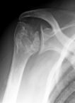
Humerol head avscular necrosis

Leg ulcers
Genito-urinary- priapism
Cerebral-stroke
Spleen- initially may cause splenomegaly due to extra medullary haemopoiesis ( due to anemia) but later splenic infarct and hyposplenism. Patients tend to have capsulated bacteria infection and Salmonella osteomyelitis.
Chest-acute chest pain
( remember that these are all due to thrombotic events)

Physical Signs
Patients tend to be pale with jaundice. Hepatomegaly may be present. Look for chronic leg ulcers.

Investigations

Full blood count- low Hb with features suggesting haemolysis such as increased reticulocyte counts
LFT- increased bilirubin and AST
Peripheral blood film- sickled cells
Haemoglobin electrophoresis- to determine variant haemoglobin
X-ray- to look for vascular necrosis if patients present with joint pain.

Management

During sickle crisis ( oxygen, analgesia, rehydration)
Long term management
- prophylactic penicillin for prevention of penumococcal infection,
- management of anemia, however be careful about secondary iron overload due to multiple transfusions
- folate supplements

Tips for MRCP

1) Always suspect Sickle Cell Disease if a patient has anemia and chronic leg ulcers.

Wednesday, November 08, 2006

Haematology in MRCP-Polycythaemia

Haematology in MRCP(1)- Polycythaemia

I am going to talk about a few important topics that are frequently asked in haematology, MRCP. The first topic that I want to talk about is polycythaemia.

Polycythaemia or erythrocytosis is defined as an increased concentration of red cells, usually with a corresponding increase in hemoglobin level.

I find the following classification of polycythaemia useful and easy to remember.


There are two main types of polycythaemia- Relative polycythamia ( pseudopolycythaemia) or absolute polycythaemia ( true polycythaemia).

Relative polycythaemia means that the absolute red cell counts are normal but due to haemoconcentration because of decreased plasma volume, patient’s haemoglobin is raised. This can be due to any kind of dehydration, however, there is a popular condition often asked in MRCP, it is an obscure condition of unknown origin known as Gaisbock’s syndrome (stress polycythaemia). It is a kind of relative polycythaemia.

For true polycythaemia, the absolute red cell counts are raised and it can be subdivided into either primary ( without obvious reason) or secondary ( with an underlying reason). For primary cause, the most important disease you must learn is polycythaemia rubra vera which I will cover in my future post. For secondary polycythaemia, the haemoglobin can be raised appropriately ( due to increase physiological need) or inappropriately.

There are three common tumours that cause secondary polycythaemia- namely renal cell carcinoma, hepatoma and hemangioblastoma ( very popular question in MRCP!). As for the examples of appropriate secondary polycythaemia, the causes are easy to remember because patients with all these conditions are hypoxia chronically, therefore they need an increase haemoglobin level to survive!

Tuesday, October 31, 2006

Cardiac Catheterization in MRCP(3)

Cardiac Catheterization in MRCP (3)

Today I am going to talk about the last part of this important topic- Cardiac Catheterization in MRCP. In my previous two posts, we have learned about some common cases concerning cardiac catherization in MRCP. I am going to show two more cases here,

Case 1:

Coarctation of the aorta


This is an easy case!There is a steep systolic gradient between the left ventricle and the femoral artery; the gradient is calculated as 190 – 150 = 40 mmHg. Therefore the diagnosis is Coarctation of the aorta.

Case 2:

Tetralogy of Fallot


You notice a combination of pressure and oxygen saturation abnormalities. The abnormalities are,

i) Step-down in oxygen saturation between LA and LV, indicating right to left shunt at the level of the ventricles, therefore there is presence of VSD.
ii) Pulmonary stenosis: there is an 89 mmHg gradient across the pulmonary valve (RV systolic – PA systolic).
iii) RVH: Right ventricular pressures are high and there is a right to left shunt, as indicated by the oxygen saturations

Hope you find this information useful. Remember, in MRCP examination, you may be given the result of a cardiac catheterization and asked about the correct physical signs of a patient. In other type of question setting, you maybe given some important physical signs and the examiners want you to find the correct cardiac catheterization results.

Monday, October 23, 2006

Poisoning in MRCP(III)

Poisoning in MRCP (III)-Tricyclic Antidepressants

The third serial of this topic- poisoning in MRCP, I am going to talk about tricyclic antidepressants. Examples of tricyclic antidepressants are amitriptyline, imipramine, doxepin etc.It is a common case scenario you would see during your internship/ housemanship. The reason is simple, doctors prescribe tricyclic antidepressants for patients with depression and these patients tend to use drugs for suicide.

As you might remember, tricyclic antidepressants help depressed patients through inhibition of reuptake of noradrenaline or serotonin in the brain, however, there are a few other effects of tricyclic antidepressants that explain its side effects,


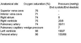
1) it has central and peripheral anticholinergic effects
2) it causes depression of cardiac contractility
3) it slows down intraventricular and artrioventricular conduction ( cardiac conduction)
4) it causes CNS toxicity such as agitation, confusion , coma and seizures


Symptoms of Toxicity

Remember that all these symptoms are due to cardiac toxicity and anticholinergic effects of tricyclic antidepressants.

Cardiac toxicity
Cardiac arrhythmias, supraventricular or ventricular arrhythmias leading to hypotension, pulmonary oedema, therefore patients may present palpitation and breathlessness

Anticholinergic toxicity ( autonomic toxicity)
Dry mouth, urinary retention, dilated pupils, constipation and hyperreflexia

CNS toxicity
Seizures ( may cause severe metabolic acidosis), coma, confusion

Treatment

Gastric lavage
Continuous cardiac monitoring is mandatory
IV Sodium Bicarbonate may be useful to maintain arterial PH if patients develop severe metabolic acidosis
DC shock may be needed, however, anti-arrhythmias are contraindicated
Fluid resuscitation if hypotension
Haemodialysis is not useful.

Tips for MRCP
1) Remember that if you see a patient with dilated pupils, confusion and cardiac arrhythmias , always consider tricyclic antidepressants.

Saturday, October 21, 2006

Rheumatology Questions in MRCP

MRCP Rheumatology Questions

Let's us take a break and look at some of common rheumatology questions in MRCP,

1) A 30 year gentleman was admitted to ward due to history of backpain for 3 months. He denied small joints pain and history of family members having the same problem. There was no history of dysuria and red eye.Below is his X-ray. What is the diagnosis?


1) Ankylosing Spondylitis
2) Riter’s syndrome
3) Psoriatic arthropathy
4) Enteropathic spondylitis
5) Seronegative Rheumatoid arthritis

2) This gentleman presents with swollen joint and fever.


What is likely to be found on microscopy of aspirated synovial fluid?

1 )Bipyramidal crystals that exhibit strong positive birefringence under polarised light
2 )Gram positive cocci in clusters
3 )Needle-shaped crystals that exhibit strong negative birefringence under polarised light
4 )Rhomboid crystals that exhibit weak positive birefringence under polarised light
5 )Small, non-birefringent crystals visible only under electron microscopy

3) A 30 year-old man is admitted to casualty with a 24 hour history of a painful and swollen right knee. He denies any previous history of joint problems. Over the last two days, he has also noticed redness and soreness in both eyes. He has returned from a business trip to Kuala Lumpur a fortnight ago.
On examination, his temperature is 38.5°C. His eyes are red. His right knee is hot, swollen and tender to palpate. No other joint appears to be affected.
Investigations:
Hb 12.9 g/dl
WBC 14.0 x 10⁹/l
Platelets 200 x 109/l
ESR 75 mm/h

Blood cultures
No growth after 48 hours

Urinalysis
No blood, glucose or protein detected

Knee x-ray
Soft tissue swelling around left knee
What is the most likely diagnosis?

1 )Gout
2 )Gonococcal arthritis
3 )Reiter's syndrome
4 )Rheumatoid arthritis
5 )Viral arthritis

4) Which of the following statements are correct regarding this patient's condition?


1 )It occurs more commonly in men
2 )Rheumatoid factor is positive in >90% of cases
3 )It is associated with an erosive arthritis
4 )Raynaud's phenomenon is a feature in ~10%
5 )It is associated with a reduced transfer factor

5) A 22 year old lady presents with typical erythema nodosum. She has a low grade fever and bilateral ankle arthritis but no other symptoms and has no medical history. There is no history of travel abroad and she is on no medication. Which of the following would be the most appropriate investigation for this patient?

1 )Barium enema
2 )Chest x-ray
3 )ESR
4 )Upper GI endoscopy
5 )Viral titres

Get your answers here!

Tuesday, October 10, 2006

Cardiac Catheterization in MRCP(2)

Cardiac Catheterization in MRCP (2)

In my previous post, I talked about a few important examples of cardiac lesion that give you abnormal oxygen saturation, today I am going to give you a few important cardiac lesions ( mainly valvular lesions ) that will give you abnormal pressure during cardiac catheterization.

Before we proceed , remember the normal cardiac pressure and oxygen saturation


Case 1: Mitral Stenosis

Look at the following cardiac catheterization result of a 40-year old lady

The diagnosis is mitral stenosis, you notice a few abnormal results here,

i) The catheter data show a gradient across the mitral valve (LA pressure – LV end diastolic pressure) .Remember that usually LA pressure equals to LV end systolic pressure; you can use the PCWP as a surrogate for LA pressure. In this case the gradient is 26-6 = 20 mmHg.
ii) There is also evidence of right ventricular hypertrophy, with markedly elevated RV pressures due to secondary pulmonary hypertension.

Case 2: Aortic Stenosis

A 65- year old lady was admitted to hospital due to syncopal attack, below is the cardiac catheterization results.

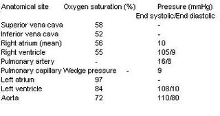
There is a systolic gradient of 81 mmHg across the aortic valve (LV systolic pressure – aortic systolic pressure), indicating severe aortic stenosis. Remember that hypertrophic cardiomyopathy also presents with a similar result, howeve, patients tend to be younger!

Case 3: Aortic regurgitation

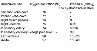
This is an easy case, you notice a wide pulse pressure ( aorta :150/40), therefore the diagnosis is aortic regurgitation.

I would talk about a few more cases in my last post.

Saturday, October 07, 2006

AIDS Defining Conditions in MRCP

AIDS Defining Conditions in MRCP-Kaposi's Sarcoma

I talked about AIDS/HIV three months ago in my blog. Today, we are going to revisit this common disease again because it is a very popular disease asked in MRCP.

There are a few simple facts to remember about HIV,

1) It is a retrovirus ( Family: Retroviridae) . HIV is completely dependent upon CD4 cells for replication and survival.
2) HIV leads to a progression fall in T-helper cells ( CD4) and a failure of T-cell proliferation.
3) HIV infection can be divided into 3 stages- acute seroconversion ( patients usually present with viral-like fever) , intermediate stage ( asymptomatic) and advanced stage ( AIDS)

Advanced stage of HIV (AIDS) is defined when the patient’s CD4 counts drop below 200 /cmm.

When our CD4 drops below 200, we are prone to get all kinds of rare infections ( opportunistic infections) which are uncommon in immune competent hosts.

There are a few AIDS defining conditions that are commonly asked in MRCP examination, these conditions are,

1) PCP
2) Toxoplasmosis
3) Cytomegalovirus
4) Kaposi’s Sarcoma
5) Cryptococcus meningitis
6) Extrapulmonary tuberculosis and atypical mycobacterium
7) Non-Hodgkin’s lymphoma


and others, however, I think these are the most important diseases you must learn.

About PCP, I think I have covered adequately in my previous post. Today I am going to talk about Kaposi’s Sarcoma (KS),

OK, this disease was considered to be very rare before the era of AIDS. It mainly affects elderly men of Mediterranean or Jewish heritage, organ transplant patients, or young adult African men. KS was named for Dr Moritz Kaposi who first described it in 1872.

In patients with AIDS, this tumuor tends to develop in the tissues below the skin surface, or in the mucous membranes of the mouth, nose, or anus. It is always described as raised blotches or lumps that may be purple, brown, or red. Sometimes the disease causes painful swelling, especially in the legs, groin area, or skin around the eyes. KS is caused by a herpes virus called Human Herpes Virus 8 (HHV-8).

However, KS is rare among Asian patients with AIDS as compared to Western patients.
In the skin, KS may not have to be treated if there are only a few lesions. Skin lesions can be:

Frozen with liquid nitrogen,
Treated with radiation,
Cut out surgically,
Injected with anti-cancer drugs or interferon alpha


A few photos of KS are shown below,


Tips for MRCP,
1) Remember that you may not only see KS occurs at skin but also mucous membrane

Tuesday, October 03, 2006

Spondyloarthropathies in MRCP (1)

Spondyloarthropathies in MRCP (1)

This is a group of disorders with following characteristics, ( remember that spondyloarthropathies are popular questions in MRCP.

1) they are seronegative ( Rheumatoid factor negative)
2) usually larger joints are involved such as knees, ankles and sacro-iliac joints
3) if peripheral joints are involved, they are usually asymmetrical
4) characteristic articular features include enthesitis ( inflammation at sites of tendon insertion),dactylitis and scaroilitis.
5) strong association with HLA-B27

The arthropathies that are under this group are,
1) Ankylosing spondylitis
2) Reiter’s syndrome
3) Psoriatic arthropathy
4) Inflammatory Bowel athropathy ( Enteropathic arthritis)

Today I am going to talk about Ankylosing Spondylitis, it is a common short case as well if you are sitting for MRCP PACES, find more discussion at PassPACES.com

Clinical Presentations

Back pain and stiffness, usually happens during the third decade
Peripheral joint pain ( less common )
Uveitis

Physical Signs

Reduced spine movement and chest expansion
‘Question mark’ posture
Anterior uveitis
Anemia of chronic disease
Aortic regurgitation murmur
Achilles tendinitis
Apical fibrosis

Investigations

Sacro-iliac joints involvement


Spine xray- loss of lumbar lordosis, Bamboo spine ( calcification in anterior and posterior spinal ligaments)
Enthesitis
HLA-B27 positive ( about 90%)

Treatment

NSAID, NSAID, NSAID + physiotherapy
Disease modifying drugs have no effect on central disease ( spine) and maybe useful in peripheral disease ( peripheral joints involvement!)

Tips for MRCP,

1) They may show a spine x-ray or pelvic xray with classical history of ankylosing spondylitis, remember how to look for Bamboo spine and sacro-iliac joint involvement!

Tuesday, September 26, 2006

Poisoning in MRCP(II)

Poisoning in MRCP (II)-Salicylates

In the second serial of this topic- poisoning in MRCP, I am going to talk about salicylates poisoning. It is an important topic in MRCP Part 1 and 2 as well as in your clinical practice. The reason is simple, salicylates can be obtained easily because it can be found in aspirin .You certainly know many patients are on aspirin if you go to ward everyday and at one time, some doctors even suggested to put aspirin in our tap water!


Before we discuss common presentations of a patient with salicylates poisoning, we must know the pathophysiology of salicylates overdose. Salicylates stimulate the respiratory centre initially and cause respiratory alkalosis. However, salicylates also interfere with carbohydrate metabolism and lead to accumulation of lactic acid and lead to metabolic acidosis.

Symptoms of Toxicity ( ASPIRIM)

Acute renal failure- symptoms of acute renal failure
Salicylism- deafness, tinnitus, vomitting
Pulmonary edema or cerebral edema (confusion)
Increased temperature
Respiratory alkalosis- hyperpnoea
GI disturbances and haemorrahge
Metabolic acidosis

Signs of toxicity

Air hunger ( due to metabolic acidosis)
Hyperpnoea ( due to respiratory alkalosis)
Remember that initially, there is respiratory alkalosis but later patient will have metabolic acidosis

Interactions

Increases anticoagulation effect
Low dose of aspirin precipitates gout

Treatment

Gastric lavage
Forced alkaline diuresis
Haemodialysis is indicated in severe cases

Hope you know how to answer your MRCP questions about salicylates after this post!

Saturday, September 23, 2006

Common Mistakes Candidates Make in MRCP

Common Mistakes Candidates Make in MRCP

OK, today I am not going talk about medicine but I would like to highlight to you a few common mistakes candidates make when preparing for thier MRCP,

1) I need at least one year or even more to prepare for MRCP Part1 and 2, but the fact is if you have a proper plan to study, you only need 5-6 months to cover every important topic.

2) I need to study a lot of books to know every fact, but the truth is you only need to stick to one good medical book. The trick is to do as many BOF as possible so that you know your weakness. Spend more time on your weak topics!

3) I need to take long leave to study their books. Medicine is always best learned beside your patients. If you see a SLE case today, go back and learn everything about SLE and find all BOFs about SLE and answer them!

4) I need to sit many times to pass the exam. You only need one attempt to pass MRCP if you prepare early and build up your confidence.

5) I must pay to get BOFs from the net, but the fact is there are a lot of sites out there which provide your free BOFs, just visit these sites and answer all these questions.

6) I must be very smart to pass MRCP, not quite, all my friends who pass their MRCP actually were not smart students when they were in medical schools.

Tuesday, September 19, 2006

Poisoning in MRCP(1)

Poisoning in MRCP(1)-Lithium

For those who recently sat for thier Part 1, good luck to all of you and hope for the best!You can do it!
" There is always hope !"

My friend sat for his MRCP Part 1 in Singapore recently. He said that questions asked in MRCP are getting more difficult to answer now. I would like to talk about poisoning today. There are a few important subtopics you must remember when learn about common drugs which are asked in MRCP.

1) Common symptoms and signs when there is poisoning.
2) Possible antidotes.
3) Drugs interactions
4) Whether the drug can be cleared by dialysis ( very important fact to remember!)

Today, I am going to talk about Lithium. If I can still remember, this drug was asked in my MRCP Part 1 in 2003.

Introduction:

Lithium is used as mood stabilizer and can be used as a treatment for acute mania/hypomania. It has a narrow therapeutic range ( <1mmol).>Symptoms for toxicity (LITHIUM!!)

- Loose motion
-Impaired vision
-Tremor
-Hypothyroidism symptoms
-Increased thirst ( polydypsia)
-Urine output increased ( polyuria)
-Muscle weakness/metallic taste

Signs for toxicity

-hyper-reflexia
- ataxia/dysarthria
-Confusion/fits

Interactions

NSAID, Thiazide, Phenothiazide, pheytoin and methyldopa increase lithium toxicity

Treatment

No specific antidotes but dialysis may be indicated!

Wednesday, September 13, 2006

Lung Cancer in MRCP

Lung cancer in MRCP

Lung cancer ( bronchial carcinoma) is a very popular topic in MRCP exam. The reason is quite simple, lung cancer is always among the top three cancer killer in males throughout the world. Cigarette is thought to be the major cause of lung cancer. There are a few important facts about lung cancer you must know,


1) There are 5 major subtypes of lung cancer

OSLAA
- Oat cell ( small cell), squamous cell, Large cell , adenocarcinoma and alveolar cell carcinoma.
Small cell lung cancer is usually the commonest type you will see in your clinical practice. Other subtypes can be put under as non-small cell lung carcinoma.

2) There are three main complications of lung cancer

The complications can be either it is due to local spread of the disease, distant metastasis or paraneoplastic syndrome. Remember that Para-neoplasic complication is frequently asked in MRCP! The most popular question asked is about Eaton-Lambert syndrome!

3) Absolute contraindications for surgical intervention are presence of distant metastasis, malignant pleural effusion, FEV1<0.8ll,other>

Clinical presentations

Classical symptoms include loss of weight, loss of appetite, cough, haemoptysis or symptoms due to underlying Para- neoplastic syndromes.

Physical signs

The physical signs can be divided according to type of complication as mentioned above.

Local spread of disease- pleural effusion, recurrent laryngeal nerve palsy ( hoarseness of voice) , superior vena cava obstruction

Distant metastasis- bony tenderness, hepatosplenomegaly

Paraneoplastic syndrome- Cushing’s syndrome, clubbing+/- hypertrophic pulmonary osteoarthropathy. (HPA)

Paraneoplastic syndrome

There are a few main presentations that you must always remember,

a) Endocrine

SIADH, Ectopic ACTH ( especially in small cell)
Hypercalcaemia- ( in squamous cell)
Carcinoid-like syndrome…… etc, etc ( Make sure your remember these four!)

b) Neurology
Remember Eaton- Lambert(EL) and how to differentiate from Myasthenia gravis.
Remember that in EL syndrome,
- usually affect proximal limbs and trunks, ocular and bulbar rarely affected
- hyporeflexia
- repeated muscle contraction may lead to increased muscle strength
( Source: Oxford Handbook of clinical medicine)


c) others- HPA, clubbing, some very rare skin lesions ( occasionally asked in MRCP PART 1 such as Erythema gyratum repens, dermatomyositis, acanthosis nigricans)

Investigations
The most important investigation- CXR but definite diagnosis may only be made after bronchoscopy, lymph node biopsy or even CT-guided biopsy.

Treatment

Small cell is usually not operable on presentation, chemotherapy may be useful
For non- small cell cancer, surgical resection if possible

Sunday, September 10, 2006

Cardiac catheterization in MRCP

Cardiac catheterization in MRCP

I found questions about cardiac catheterization are always very challenging in MRCP Part 2 exam. When I sat for my Part 2 three years ago, they would usually show you the pressure and saturation from a cardiac catheterization and ask you what the underlying cardiac lesion is, however, I notice that recently they have switched the way of asking this type of question. They prefer to give you a case with important physical signs and ask you to choose the correct cardiac catheterization results.

Anyway, start form basic first, remember the normal and pressure of each cardiac chamber as below,

Left heart
Aorta-------120/80 ( pressure)---------98% (saturation)
LV----------150/5-10------------------98%
LA--------------------------------------98%

Right heart
RA------------0-8 (mean pressure)-----74%
RV-----------15-30/0-8----------------74%
PA-----------15-30/3-12---------------74%
SVC-------------------------------------74%
IVC-------------------------------------70%
PCWP--------1-10 ( mean)

There are two main groups of questions in MRCP, either a saturation or pressure problem, we would talk about saturation problem first today.
I would give you a few examples and explain to you the underlying cardiac lesions,

Case 1:

---------------Pressure mmHg --Saturation%
Right heart
SVC ------------------5-------------- 72
RA -------------------6-------------- 76
RV----------------- 25/0-5 ----------76
PA -----------------25/10----------- 77

Left heart
LV ---------------140/0-12---------- 97
Aorta --------------140/75 -----------97

You notice that the saturation at the superior vena cava is 72% and increases to 76%, if you remember your anatomy, blood from SVC would go to right atrium (RA) and the saturation should be the same ( see above image again), therefore , there must be presence of mixture of blood from more oxygenated blood from left atrium and right atrium. Yes, you are right, the diagnosis is atrial septal defect!

Case 2:

----------------Pressure (mmHg) ----Oxygen saturation (%)
SVC--------------- - ---------------------72
RA ------------------7--------------------72
RV----------------- 50/12----------------86
PCWP-------------- 16 ------------------- -
LV -----------------90/12----------------96
Aorta ---------------100/50---------------97

This question is easy, you notice there is a sudden increase in oxygen saturation from RA to RV, therefore, there must be mixture of blood over RV and LV, the diagnosis is Ventricular septal defect( VSD).

Case 3:

-------------------Pressure (mmHg)------ Oxygen saturation (%)
IVC -------------------------------------------60
RA ---------------------20---------------------66
RV--------------------- 100/20---------------- 67
PA ---------------------100/30---------------- 67
LV ----------------------100/10 ---------------70
Aorta-------------------- 105/80---------------70

This case is more complicated, however, stay calm, apply basic principles. You notice that there is an increase of oxygen saturation form IVC to RA ( sounds like it is an atrial septal defect). However, you notice that RV’s pressure is high and even equals to that of LV’s. Another abnormality you notice is the saturation of LV and aorta is low ( as compared to more than 95% in normal subjects). So…… the diagnosis is Eisenmenger’s syndrome secondary to ASD.

Case 4:

----------------------------Pressure (mmHg) --------Oxygen saturation (%)
Superior vena cava ----------------------------------------77
RA -----------------------------6--------------------------78
RV -------------------------------------------------------- 78
PA ---------------------------50/20----------------------- 86
PCWP--------------------------16-----------------------------
LV--------------------------120/11------------------------96
Aorta------------------------- 130/60----------------------97

This case is simple, you notice there is high pressure present over Pulmonary artery with sudden increase in oxygen saturation, therefore mixture of blood must be present over the pulmonary artery. Yes, you are right, you are dealing with Patent Ductus arteriousus.

Ok, that is enough for today, we have learn about cardiac lesions that give you abnormal oxygen saturation during cardiac catheterization, I would talk more about valvular heart lesions that give your abnormal pressure during cardiac catheterization in future posts.

Friday, September 08, 2006

Paget’s disease of bone in MRCP

Paget's disease of bone

Disease of unknown origin. There is increased bone turnover due to an increase in bone osteoclast activity , which leads to increased osteoblast activity. Increased bone remodeling leads to bone enlargement, deformity and weakness.


Clinical presentations

Usually bone pain, bone deformity and deafness

Physical signs

You may notice bone deformity and warm bones. Rare in patients less than 40 years old. Rare as well in tropical countries.

Investigations

I notice that a lot of candidates have problems interpreting Calcium, phosphate, ALP in various conditions, I find this the following table useful,

Bone disease calcium Phosphate ALP
Paget’s Normal Normal ^^
Myeloma ^/Normal Normal unless fracture
Osteomalacia down down ^
Osteoporosis Normal Normal normal
Bone metastasis ^ ^/ normal ^

( It is even more confusing for hyperparathyroidism, I would cover that in future post)

Remember that level of ALP reflects osteoblast activity, therefore, if there is no new bone formation, ALP level would not be raised. This also explains ALP is high in growing children! On the other hand, level of urinary hydroxyproline reflects osteoclast activity, when there is breakdown of bone, the level would be raised.

Diagnosis

Based on typical x-ray appearance and raised AP with normal Calcium and phosphate

Coarse trabeculation and bony expansion

Complications

Nerve/ cord compression
Osteogenic sarcoma
Heart failure ( high output)

Treatment

Bisphosphonates is the treatment of choice. It inhibits bone resorption ( osteoclast activity) . It is also used in management of hypercalciamia especially in bone metastasis and osteporosis!

Tips for MRCP

Remember that patients with Paget's disease always have a raised ALP with normal calcium and phosphate !

Sunday, September 03, 2006

Genetics in MRCP

Today I am going to tell you a few genetics conditions which are commonly asked in your MRCP PART 1.

1) Autosomal dominant inheritance
- generally ‘ structural-type’ disorders

a) adult polycystic kidney disease
b) myotonic dystrophy
c) Elers-Danlos and Marfan’s syndrome
d) Hereditary haemorrhagic telangiectasia
e) Huntingtons chorea
f) Intestinal polyposis
g) Neurofibromatosis
h) Otesogenesis imperfecta
i) Tuberous sclerosis

2) Autosomal Recessive inheritance
- generally ‘metabolic type’ disorders

a) Infantile polycystic kidney
b) Alfa1-antitrpsin deficiency
c) Cystic fibrosis
d) Most inborn errors of metabolism ( galactosaemia, glycogen storage diseases etc)
e) Haemoglonbinopathies ( sickle cell disease and thalassaemias)
f) Wilson’s disease
g) Friedreich’s ataxia

3) Sex-linked dominant inheritance

a) Vitamin D risistnact rickets

4) Sex-linked recessive inheritance

a) G6PD deficiency
b) Haemophilia A and B
c) Lesch-Nyhan syndrome
d) Immunodeficinecies- agammagobulinaemia

However, there are a few types of muscular dystrophies you should remember,

Duchenne - X linked recessive
Becker -X linked recessive
Limb girdle -Autosomal recessive
Facio-scapulo-humerol -Autosomal dominant

Tips for MRCP

Remember how to interpret a family tree, it is the commonest way how genetics of above disoredrs are asked in MRCP!

Thursday, August 31, 2006

Gout in MRCP

GOUT IN MRCP

I saw this patient during my clinic follow up recently, so I am going to discuss with you about gout today. Gout is commoner than you think. Gout is due to deposition of uric acid crystals in joints. Certain joints are commonly involved such as first toe, ankle and small joints of hands.


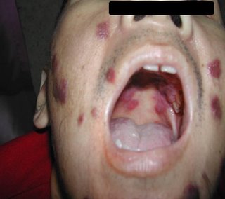
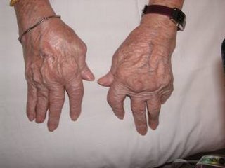
Chronic tophaceous gout with tophi!

Clinical Presentations

There are a few possible manifestations of gout. These are,
a) asymptomatic hyperuricemia
b) acute arthritis
c) chronic arthritis
d) chronic tophaceous gout

Physical signs

You may find swollen and tender joint if patient has acute arthritis, you must always consider septic arthritis as your differential diagnosis. Patient may also present to you with chronic tophaceous gout as I have shown you as the images above.

Precipitating Factors for Acute Attack

There are a few precipitating factors for you to get acute attack of gout. These factors are,
a) Trauma
b) Drugs such as diuretics, aspirin ( always a popular question in MRCP, remember hat only low dose of aspirin precipitates gout)
c) Copious consumption of alcohol
d) Dehydration
e) Surgery
f) Infection
g) Food high in Purines
h) Induction chemotherapy for certain cancers such as leukemia

Treatment

Pharmacological and non-pharmacological treatments are available.
a) Diet and lifestyle changes
b) Drugs such as Allupurinol, Probenecid. Remember that allupurinol interacts with Azathioprine and cyclophosphamide and increases the toxicity of these cytotoxic drugs ( VERY popular question in MRCP!)

Gout Vs Pseudogout

Remember that pseudogout is acute arthritis resulting from the release of calcium pyrophosphate ( deposited in the bone and cartilage) into the synovial fluid.
Gout /Pseudogout

More severe and short lasting / less severe and longer lasting
Usually first toe involved/ mainly the knee
Negatively birefringent/ Positive
Needle shape crystal / Rhamboishape
No calcium deposition / Ca deposition on X ray

Tips for MRCP

1) Remember that allupurinol also can cause Steven Johnson syndrome!
2) Related post, click here!

Wednesday, August 30, 2006

Popular Drugs in MRCP (7)

Popular Drugs in MRCP- Methotrexate

Today I am going to discuss with you with another popular drug in MRCP. Methotrexate (MTX) is a folic antagonist and also a dihydrofolate reductase inhibitor with potent immunosuppressive activity.

MTX is used in a lot of autoimmune diseases such as Rheumatoid arthritis , vasculitis such as Wegener’s granulomatosis. Certain types of cancer and even in ectopic pregnancy!

Common side effects

Nausea, vomiting
Mucosal ulcer
Hepatotoxicity
Lung fibrosis

Remember that other drugs that can cause lung fibrosis are amiodarone,bulsuphan, bleomycin, nitrofurantoin, hydralazine.

Tips for MRCP

1) Side effect of MTX that is commonly asked in MRCP is lung fibrosis and hepatotoxicity.

Tuesday, August 29, 2006

Good Luck To All!

Good Luck!

Hi, For those who are sitting for their MRCP (UK) and MRCP (Ireland) Part 1 soon, good luck to all of you! You can do it, just remember to have a good sleep before your exam and stay calm in the examination hall!

Saturday, August 26, 2006

Rheumatoid Arthritis in MRCP

RHEUMATOID ARTHRITIS

RA is a type of autoimmune disease mainly involving joints. It leads to chronic inflammation of joints. However, candidates must remember that it is a multi-system disease that might involve other organs as well such as skin, eye , lung and cadiovascular. More female are affected with a ratio of 3:1.

Clinical Presentations

Majority of patients present with small joints pain. However, patients may have systemic symptoms such as fever, weight loss and fatigue.
Some patients may have eye symptoms such as red , painful eye ( due to scleritis and episcleritis)
There is possibility of lung fibrosis
Patients may have neurological deficits due to alanto-axial subluxation or mononeuritis multiplex.

Physical signs

Classical hand deformities are Z deformity, Swan neck , Boutonniere deformity, to learn more click www.passpaces.com/issue1.html
Lung fibrosis


Diagnosis

Diagnosis can be made based on American Rheumatism Association Criteria:
1) Morning stiffness >1 hour for 6 weeks or more
2) Swelling of at least 3 joints for 6 weeks or more
3) Swelling of wrist, MCP,PIP joints for 6 weeks or more
4) Symmetrical joints pain for 6 weeks or more
5) Subcutaneous nodules
6) +ve RF
7) Classical X ray appearance ( periarticular osteopenia)

Four or more out of seven criteria above, the diagnosis can be made

Investigations

Rheumatoid factor (RF)- however this is not specific, it can be positive in normal population ( false postive)
FBC- anaemia- Remember 4 causes of anaemia in RA- Chronic illness, Felty syndrome, Drug-induced (anaemia due to UGIB, secondary to gold, methotraxate)
Xray- lung ( fibrosis), hand, cervical ( subluxation)

Treatment

Disease modifying drugs such as sulphasalazine, methotraxate, gold
Latest drugs such as etanercept and infliximab- popular questions in MRCP, click here to learn more!

Tips for MRCP

1) Remember the side effects of drugs used to treat RA including etanercept and infliximab!

Friday, August 25, 2006

Sarcoidosis in MRCP

SARCOIDOSIS

Sarcoidosis is always a popular question in respiratory section in MRCP. It was first described by a London surgeon-dermatologist, Dr. Jonathan Hutchinson in 1877. The doctor described the findings of a 50 year-old man who had large purple skin plaques on the hands and feet and a 64-year-old woman with large purple patches on her face and arms. You must remember that Sarcoidosis is a multisystem disease with unknown origin, however, in MRCP, two popular systems which are frequently asked are skin and lung.

Clinical Presentations

Usually asymptomatic, however patient may have constitutional symptoms such as fever, malaise and weight loss. Majority of patients have respiratory symptoms such as cough and shortness of breath.
Popular skin condition ( always asked in MRCP) associated with sarcoidosis is Erythema nodusom. Pateints may have arthralgia or bone pain.
Eyes-painful eyes, dry eyes
Other-dry mouth, hepatomegaly

Investigations

They would usually show show you a CXR in MRCP
Remember that 90% of CXRs have bilateral hilar LN enlargement. More advanced case may have diffuse fibrosis!


ESR-raised
Serum Calcium-raised
Serum ACE-raised
Lung function test may show obstructive changes

Diagnosis

Diagnosis can be made by lung biopsy-granuloma! Remember that serum ACE is not specific nor sensitive.

Treatment

The main treatment for sarcoidosis is prednisone. Prednisone is a corticosteroid, or anti-inflammatory drug. Sometimes it is used with other drugs. Sometimes other corticosteroids are used.

Tips for MRCP

1) If you are asked about a patient with painful skin lesions over shin area with dry cough, always think of sarcoidosis.

2) A patient with high calcium level and dry cough , think of sarcoidosis although there is possibility of lung cancer with bone metastasis!

Monday, August 21, 2006

MRCP Part 1-Mock Exam (2)

MRCP Part 1 Questions

Here are some questions for MRCP Part 1, take the test and look at the answers later!

1)Ostium secundum ASD is associated with
A tricuspid regurgitation
B left bundle branch block
C fixed splitting of the second heart sound
D onset of atrial fibrillation in the second decade
E early onset of heart failure in the second decade

2)A 34 year old male presents with episodes of breathlessness on exertion. Examination reveals a loud P2 and fixed splitting of the second sound. Which of the following may be responsible for these signs?
1 ) Maternal chicken pox infection 2 ) Maternal thalidomide therapy
3 ) 47 XXY karyotype 4 ) Homocystinuria 5 ) Excess maternal alcohol consumption

3)A 35-year-old healthy woman has a faint systolic murmur on physical examination. An echocardiogram is performed, and she is found to have a bicuspid aortic valve. In explaining the meaning of this finding to her, the most appropriate statement is that?

1 ) An aortic valve replacement is eventually likely to be required.
2 ) Other family members are likely to have the same condition
3 ) She should be treated with a cholesterol-lowering agent
4 ) The problem resulted from past injection drug usage
5 ) This is one manifestation of an underlying autoimmune disease process

4) A 24-year-old woman develops infective endocarditis involving the aortic valve. She receives a porcine bioprosthesis because of her desire to have children and not to take anticoagulantmedication. After ten years, she must have this prosthetic valve replaced. Which of the following pathologic findings in the bioprosthesis has most likely led to the need for replacement?
1 ) Calcification with stenosis
2 ) Dehiscence
3 ) Infective endocarditis
4 ) Strut failure
5 ) Thrombosis

5) The following can cause bradycardia: EXCEPT
A hypothermia B hypothyroidism C severe anaemia D subdural haematoma E shock

6)The following are recognised causes of reversible dilated cardiomyopathy:- EXCEPT
A alcohol B Selenium deficiency C Acromegaly D Lead poisoning E Coxsackie virus

7) Which of the following concerning congenital heart disease is correct?
1 ) ASD is the commonest malformation at birth
2 ) congenital complete heart block is usually associated with Anti-Ro antibodies in the mother 3 ) Ebstein's anomaly is associated with maternal exposure to lithium carbonate
4 ) Hypoplastic left heart syndrome is characterised by a large, dilated left ventricle
5 ) Osteogenesis imperfecta is associated with aortic stenosis

8) Coarctation of the aorta is: EXCEPT
A usually congenital but may be aquired
B recognised by absent or delayed femoral pulses
C a common cause of heart failure in infancy but an uncommon cause of hypertension in adults D associated with an increased incidence of an aortic bicuspid valve
E a cause of left to right shunting of blood

9)A 65-year-old woman, a heavy smoker for many years, has had worsening dyspnoea for the past 5 years, without a significant cough. A chest X-ray shows increased lung size along with flattening of the diaphragms, consistent with emphysema. Over the next several years she develops worsening peripheral oedema. BP 115/70 mmHg. Which of the following cardiac findings is most likely to be present?
1 ) Constrictive pericarditis 2 ) Left ventricular aneurysm 3 ) Mitral valve stenosis
4 ) Non-bacterial thrombotic endocarditis 5 ) Right ventricular hypertrophy

10)Increased pulmonary vascular markings on chest x-ray are a recognized feature of:
A Pulmonary stenosis
B Mitral stenosis
C Persistent ductus arteriosus
D Primary pulmonary hypertension
E chronic constrictive pericarditis

11) A 59-year-old man who was active all his life develops sudden severe anterior chest pain that radiates to his back. Within minutes, he is unconscious. He has a history of hypertension, but a recent treadmill test had revealed no evidence for cardiac disease. Which of the following is the most likely diagnosis?
1 ) Acute myocardial infarction
2 ) Group A streptococcal infection
3 ) Pulmonary embolus
4 ) Right middle cerebral artery embolus
5 ) Tear in the aortic intima

12)Low T waves on an ECG are seen in:
A hyperkalaemia B hypercalcaemia C athletes D pericardial effusion E myelodysplasia

13)A 56 year old male with left ventricular systolic dysfunction was dyspnoeic on climbing stairs but not at rest. The patient was commenced on ramipril and frusemide.
Which one of the following drugs would improve the patient's prognosis?
1 ) Amiodarone 2 ) Amlodipine 3 ) Bisoprolol 4 ) Digoxin 5 ) Nitrate therapy

14) A 70-year-old male is referred by his GP for management of recently diagnosed congestive heart failure. The patient has a history of poorly controlled hypertension. Over the last three months he has been aware of deteriorating shortness of breath, fatigue, and orthopnea. Over the last month he had been commenced on Digoxin (62.5 micrograms daily), Frusemide (80 mg daily), and amiloride 10 mg.
On examination he has a pulse of 96 bpm regular, a blood pressure of 132/88 mmHg. His JVP was not raised, he had some scattered bibasal crackles on auscultation with a displaced apex beat in the anterior axillary line, 6th intercostal space. Auscultation of the heart revealed no murmurs and he had peripheral oedema to the mid tibia.
Investigations showed: electrolytes normal serum urea concentration 17 mmol/l (NR 2-8 mmol/l) creatinine 175 micromol/l (NR 55-110) Serum digoxin 0.7 ng/mL {therapeutic: 1.0-2.0}
One month previously his urea had been 11 mmol/l and creatinine 110 micromol/l. An ECG reveals left ventricular hypertrophy and Chest X-ray shows cardiomegaly and calcified aorta.
What is the most appropriate next step in management?
1 ) Add an ACE inhibitor to the current regimen
2 ) Add atenolol at a dose of 25mg daily
3 ) Increase digoxin to 0.25 mg daily
4 ) Increase frusemide to 80 mg twice daily
5 ) Maintain on current therapy.

15)A 17-year-old woman loses consciousness while out jogging one afternoon, as she has done for many years. She is taken to Accident and Emergency, where a chest X-ray, CT brain scan, FBC, and biochemistry are all normal. Over the next year, she develops mild dyspnea and fatigue. There are several episodes of pre-syncope. After another syncopal episode, she is referred to a cardiologist who orders and ECG that shows changes of left ventricular hypertrophy and broad Q waves. An echocardiogram reveals left ventricular and septal hypertrophy, small left ventricle, and reduced septal excursion. The septum has a "ground glass" appearance. She then dies suddenly and unexpectedly. The microscopic appearance of the septum with trichrome stain reveals myofiber disarray. Which of the following conditions is she most likely to have had?

1 ) Diabetes mellitus 2 ) Hypertrophic cardiomyopathy 3 ) Rheumatic heart disease 4 ) Systemic lupus erythematosus 5 ) Viral myocarditis

16) A 78 year old female is referred by her GP with high blood pressure. Over the last three months her blood pressure is noted to be around 180/80 mmHg. She has a body mass index of 25.5kg/m2, is a non-smoker.There are no features to suggest a secondary cause for her hypertension. Which of the following is the most appropriate treatment for her blood pressure?

1 ) Alpha-Blocker 2 ) Angiotensin Converting Enzyme (ACE) Inhibitor 3 ) Angiotensin Blocker 4 ) Beta-blocker 5 ) Calcium channel blocker

17) Infective endocarditis rarely occurs with:
A mitral valve prolapse B patent ductus arteriosus C bicuspid aortic valve D atrial septal defect E mitral stenosis

18) A 60-year-old man presents with an inferior MI and receives thrombolysis. 4 hours following initial presentation he becomes acutely breathless. His ECG demonstrates sinus tachycardia (rate 108bpm) with T wave inversion inferiorly. His ST segments are normal. On examination his JVP is elevated at 5 cm. Chest was clear to auscultation. Following 80 mg of Frusemide he deteriorates. His BP is now 80/60 and his urine output over the last 2 hours is 5 mls. What is the best investigative measure?

1 ) Arterial Blood Gases 2 ) Central Venous Pressure Monitoring 3 ) Chest X-Ray 4 ) Echocardiography 5 ) Pulmonary Capillary Wedge Pressure Monitoring

19) The following are true regarding mitral stenosis:
A it is not tolerated well in pregnancy
B there is characteristically a low wedge pressure
C in AF, the opening snap disappears
D loud murmurs if valve is high calcified
E Doppler U/S is usually inaccurate in determining severity

20) The following are recognised features of pulmonary embolism: EXCEPT

A long PR interval on the electrocardiogram
B decreased left atrial pressure
C pulmonary hypertension
D collapse of the affected lung segments
E necrosis of lung tissue

Find your answers here!

Sunday, August 20, 2006

Primary Biliary Cirrhosis in MRCP

Primary Biliary Cirrhosis (PBC)

PBC is an autoimmune disease. It is characterized by progressive inflammmation and destruction of of the small bile ducts within the liver. In the long run, it can lead to liver cirrhosis and PBC is one of the important causes of liver cirrhosis among female patients. ( 90% of PBC patients are female!)

Clinical Presentations
Usually asymptomatic but later due to biliary obstruction, patients tend to have pruritus and jaundice. Patients may present late with symptoms of liver cirrhosis such as upper GI bleeding or abdominal distention ( ascites)

Physical signs
Hepatosplenomegaly, Jaundice, scratch marks, xanthelasma, clubbing or other signs suggesting associated autoimmune diseases. Click here to see xanthelasma!

Associations
Autoimmune diseases such as Sicca syndrome, thyroiditis, RA, SLE

Investigations
I think the single test that can give a clue of PBC is LFT- you would notice markedly high ALP with raised bilirubin, IgM may be raised.
ANA may be positive
Diagnosis can be made by detecting antimitochondrial antibody. Specificity of the M2 subtype is 95-99%
Liver biopsy shows granulomas

Treatment
symtomatic relief of pruritus by cholestyramine.
Ursodeoxycholic acid may be useful
Cochicine may be used to slow down the progression
Immunosuppresants such as steroid, methotrexate may be useful
The only cure- liver transplantation

Tips for MRCP
1) Remember that if a lady has jaundice and very high ALP with pruritus, suspect PBC

Thursday, August 17, 2006

Tuberous Sclerosis

Tuberous Sclerosis in MRCP

There are a few conditions that are commonly asked in MRCP Part 1 and 2. These conditions include Tuberous sclerosis (TS), Neurofibromatosis..... etc. I am going to talk about TS today.
TS was discovered in the 1880's by a French physician named Bourneville. Remember that it is an autosomal dominant disease!

There are a few important features of TS, these include,

Facial angiofibroma
periungual fibromas
Hypopigmented macules
Shagreen patch (connective tissue nevus)
Multiple retinal nodular hamartomas
Subependymal nodule ( can be picked up in CT scan as calcification)
Subependymal giant cell astrocytoma
Cardiac rhabdomyoma, single or multiple
Lymphangiomyomatosis
Renal angiomyolipoma

Questions commonly asked in MRCP are photo-based questions, remember these photos.....
1) Periventricular calcification

2) Clinical features of TS


See more photos at www.passpaces.com

Tips for MRCP

1) A patient with recurrent seizure and CT scan shows calcification. Remember TS!

Wednesday, August 16, 2006

Popular Drugs in MRCP (6)

Rosiglitazone

Rosiglitazone is a popular drug asked in MRCP examination. It is an aanti-diabetic drug from the thiazolidinedione class. Its mechanism of action is by activation of the intracellular receptor class of the peroxisome proliferator-activated receptors (PPARs), Rosiglitazone is ofter referred as 'insulin sensitizer' because it makes the body cells become more sensitive to insulin and remove more glucose from blood.

Indication for Rosiglitazone

Type 2 Diabetes mellitus. It is usually used alone or combined with metformin

Common side effects

It can cause mild to moderate oedema and should be avoided in acute heart failure or severe heart failure patients.
Possibility of liver impairment
Diarrhoe
Headache
Hypo or hyperglycaemia

Common Interaction

Gemfibrozil increases the concentration of rosiglitazone in the blood by reducing its breakdown. Therefore, gemfibrozil may increase the side effects of Rosiglitazone!

Tips for MRCP:
1) Remember about the side effects of Rosiglitazone.

Monday, August 14, 2006

Multiple Myeloma

Multiple Myeloma in MRCP

Multiple Myeloma (MM) is a very important disease in haematology, MRCP. It is frequently asked and there are various clinical presentations in MM. Candidates must know that the malignant cells ( proliferation of plasma cells) are from bone marrow and not cortex of the bone ( as compared to osteosarcoma or scondary deposits of distant tumour to boen cortex). The monoclonal plasma cell line produces immunoglobulin which is abnormal and leads to suppression of normal immunity.

Clinical Presentations

Remember this mnemonic CRAB
Calcium abnormality - increased calcium level due to enhanced osteoclastic activity. Patients can present with confusion, dehydration, polyuria or even abdominal pain.
Renal impairment. Patients can present with tiredness
Anaemia ( due to suppression of bone marrow)- symptoms of anaemia
Bony abnormality- lytic lesions in almost 60-70%- bony pain

Physical Signs

Usually no hepatosplenomegaly but you may notice pallor, dehydration or bony tenderness.

Investigations

Monoclonal gammopathy ( usually Ig G- as compared to Waldenstrom's Macroglubinaemia which has increased Ig M)
Bone Marrow shows more than 30% plasma cells
Skeletal survey shows mutiple osteolytic lesions ( Candidates must remember that ALP of patients may be normal except there is presence of bone fracture)
ESR usually very high

Treatment

Thalidomide- Remember this drug in your medical student books? Thsi drug was usd extensively many years ago for insomnia and morning sickness for pregnant mothers but it was later found to be teratogenic. ( Remember your medical books with photo of babies without upper and lower limbs?)
However, current trials show it effectiveness against MM. Click here to learn more!

The latest drug- bortezomib!

Tips for MRCP
1) Suspect MM if patient has anaemia, renal impairment, high ESR and Calcium level.

Friday, August 11, 2006

MRCP Part 1 Questions (2)

Hope you find these neurology questions useful..........

1) A 26-year-old male presents with 2 days history of diplopia and unsteadiness. 2 weeks ago he suffered from viral fever. Examination reviews that there is complete opthalmoplegia, areflexia and gait ataxia. Which of the following blood tests is the most likely to confirm the diagnosis?
1 ) Acetylcholine receptors antibodies
2 ) Anti GM1 antibodies
3 ) Anti GQib antibodies
4 ) Anti Topoisomerase antibodies
5 ) Anti purkinje cell antibodies

ANSWER: 3

2) A 40-year-old woman is referred with a two-week history of difficulty walking . On examination, there was distal limb weakness and the power is 3/5. Tendon reflexe was absent over ankle and the plantar responses were flexor.There was no sensory loss. What is the most likely diagnosis?
1 ) polymyositis
2 ) cervical cord compression
3 ) Guillain-Barré syndrome
4 ) myasthenia gravis
5 ) poliomyelitis

ANSWER: 3

3) A 50 year old female is admitted with progressive weakness following a flu-like illness. Which of the following would exclude Guillain-Barre Syndrome as the diagnosis?
1 ) Autonomic dysfunction
2 ) Elevated protein on CSF examination
3 ) Evidence of muscle wasting
4 ) Ophthalmoplegia
5 ) Sensory involvement

ANSWER: 5

4) A 15 year old girl presents with Guillain-Barre syndrome. Her weakness continues to worsen after admission to hospital and she complaines of shortness of breath. Which of the following should be used to monitor her?
1 ) arterial blood gases
2 ) chest expansion size
3 ) FEV1/FVC ratio
4 ) PEFR
5 ) vital capacity

ANSWER: 5

5) Which of the following clinical manifestations suggests Guillain Barré Syndrome?
1 ) Weakness beginning in the arms
2 ) Asymmetrical involvement of distal muscles
3 ) Bulbar involvement in about 50% of cases
4 ) Brisk tendon reflexes
5 ) Normal CSF protein

ANSWER: 3

6)A 43-year-old woman develops a progressive, ascending motor weakness over several days. She is hospitalized and requires intubation with mechanical ventilation. She is afebrile. A lumbar puncture is performed with normal opening pressure and yields clear, colorless CSF with normal glucose, increased protein, and cell count of 5/microliter, all lymphocytes. She gradually recovers over the next month. Which of the following conditions most likely preceded the onset of her illness?
1 ) Ketoacidosis
2 ) Staphylococcus aureus septicemia
3 ) Systemic lupus erythematosus
4 ) Viral pneumonia
5 ) Vitamin B12 deficiency

ANSWER: 4

7)Common features of normal pressure hydrocephalus are EXCEPT:
A papilloedema
B The opening pressure for lumbar puncture is normal
C gait apraxia
D incontinence
E cognitive impairment

ANSWER: B

8)A 60 year-old man presents with a 2 month history of progressive confusion, gait disturbance, and urinary incontinence. Examination reveals gait ataxia. CT brain done is as follow, lumbar puncture reveals normal CSF pressure and constituents. Which one of the following managements steps is likely to be most helpful?


1 ) CSF drainage via repeated lumbar puncture
2 ) EEG
3 ) Intracranial pressure monitoring
4 ) MRI brainstem
5 ) Serum B12 and folate levels
ANSWER: 1

9)A 75-year-old man presented with an unsteady gait. He was noted to be becoming impaired with his memory and agitated at nights. His GP started an antidepressant. He was incontinent of urine. He was a heavy smoker and had lost 2 stones in weight over 2 months. His blood sugar was 10 mmol/l.
Which is the next best investigation?
1 ) CT Head
2 ) CXR
3 ) Arterial Blood gas
4 ) Thyroid function test
5 ) Blood Calcium level

ANSWER: 1

Wednesday, August 09, 2006

Guillain-Barré Syndrome

Guillain-Barré Syndrome in MRCP

This is a popular disease in MRCP part 1 and 2. In part 2, question about GB syndrome would be asked based on CSF interpretation.
GB is an acute polyneuropathy mainly affecting motor neuron. It is usually demyelinating type. Patient usually presents after recent viral/bacteria infections.

Trigerring Agents

Campylobacter jejuni ( usually cause diarrhoe)
CMV
EBV
HIV
mycoplasma species

Clinical Presentations
Acute onset of leg weakness and ascends to the upeer limbs. Peripheral numbness is possible.

Physical Signs
Lower motor lesion of both lower limbs ( sensory loss is distal and minimal)
Papilloedema
Autonomic dysfunction ( may lead to cardiac arrhytmia and death)
Cranial nerves involvement ( Miller Fisher variant- ophtalmoplegia with ataxia)

Investigations
For Miller Fisher, autoantibody GQ1B can be present.
CSF ( Cerebral spinal fluid ) shows high protein
Nerve conduction shows demyelinating pattern
Remember that to monitor patient ( in case of respiratory muscles involvement), you should monitorthe FVC ( spirometry) NOT Peak Expiratory Flow Rate (PEFR)

Treatment
IV Immunoglobulin. Plasma exchange can be considered.

Tips for MRCP:
1)If the weakness lasts longer than months, always consider CIDP ( chronic inflammatory demyelinating polyneuropathy.)
2) Major cause of death in patients with GBS is due to autonomic dysfunction and cardiac arrhytmia!

Tuesday, August 08, 2006

Journey to MRCP

I came across this saying while surfing the net.....


Success is a journey, not a destination. The doing is often more important than the outcome.
Hope that candidates who did not make it for the recent Part 1 and 2 know that even most your consultants have to struggle a few times to pass their MRCP!

Monday, August 07, 2006

Normal Pressure Hydrocephalus

Normal Pressure Hydrocephalus in MRCP

Normal pressure hydrocephalus (NPH) is hydrcephalus with normal intracranial pressure. You must remember that majority of patient has no identifiable cause for NPH. It usually happens in elderly patients.

Clinical Presentations

Triad of GUD
Gait disturbances, Urinary incontinence and Dementia.

Investigations


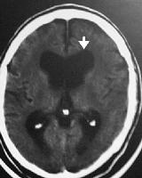
Picture source: e-medicine (arrow- dilated ventricles)

All investigations would be normal except CT/MRI head show non-obstructive hydrocephalus. PET scan mayshow hypometabolism.

Treatment

Ventricular shunting or lumbar puncture drainage of CSF

Tips for MRCP

1) Popular questions in MRCP are interpretation of CT brain, an elderly demented patient with urinary incontinence.
2) Remember that NPH is one of the important differential diagnosis of dementia!

Sunday, August 06, 2006

Autoimmune Hepatitis

Autoimmune Hepatitis in MRCP

I must tell you that this is a very important topic in MRCP gastroenterology. Besides that, it is rather common in your dily practice if you are attached to the gastroenterology unit. It is an autoimmunedisease with unknown aetiology. Usually questiosn about autoimmune hepatitis would be asked in Part 2and you are expected to know how to interpret liver function test and autoantibody profiles.Patients are usually young ladies.

Clinical Presentations

Patients usually come to you with jaundice and right hypochondrium pain. One important pointto remember about autoimmune hepatitis is that it is commonly associated with other immune diseasesuch as Sjoren's syndrome, Renal Tubular acidosis etc...

Clinical Signs

Patients may have chronic stigmata of chronic liver disease. You may find hepatosplenomegaly. If patient is on treatment, then you would find signs suggesting Cushing's syndrome. Patient mayend up with liver cirhosis and features of hypersplenism.

Investigations


Liver function test reveals increased bilirubin and ALT levels. Autoantibodies that may be positive include ANA ( up to 79-80%), Anti-smooth muscle antibodies, Anti-LKM-1 antibodies and anti-mitochondrial anti-bodies ( rarer in autoimmune hepatitis, you find this more common in primary biliary cirrhosis). Liver biopsy shows PIECEMEAL NECROSIS ( as above slaid)

Treatment

Steroid and steroid sparing agent such as Azathioprine, LIVER TRANSPLANT

Saturday, August 05, 2006

Atypical Pneumonia

Atypical Pneumonia in MRCP

Atypical pneumonia is a popular question in MRCP,a lot of candidates ask me why we call Legionella and Mycoplasma pneumonia, atypical pneumonia and why they are so important in exam? There are a few reasons,

1) These two types of pneumonia usually present with extra-pulmonary symptoms such as diarrhoe andvomitting.Besides that, they may lead to extra-pulmonary complications.
2) It is suprising common and may account up to 25% of total community acquired pneumonia. Click here to learn more about pneumonia!

A few important points to remember about these two types of pneumonia

1) Legionella pneumophilia is commonly found in cooling system ( air-conditioning system) and water carrying system.
2) Mycoplasma penumonia can cause haemolytic anemia ( cold agglutinin presents in 50%) and may lead to skin rash ( erythema multiforme)
3) Both can lead to a lot of extra-pulmonary complications such as myocarditis, pericarditis, pancraetitis, meningitis etc

Dignosis of a atypical penumonia

CXR may be non specific but remember for Legionella Pneumonia, diagnosis can be made with Legionella serology and urinary antigen test. For mycoplasma pneumonia, send for mycoplasma serology but cold agglutinin test may be helpful.

Treatment

Always remember that either macrolides or tetracycline can be used. However, add rifampicin in severe Legionella pneumonia

Tuesday, August 01, 2006

MRCP Part 1 Questions (1)

Hope you find the following questions useful,

1)A 50-year-old male presented with acute respiratory failure during an episode of fulminant sepsis and was thought to have developed adult respiratory distress syndrome (ARDS). Which of the following would support a diagnosis of ARDS?

1 ) High pulmonary capillary wedge pressure
2 ) High protein pulmonary oedema
3 ) Hypercapnia
4 ) Increased lung compliance
5 ) Normal chest X-ray

ANSWER: 2

2)In malignant hyperpyrexia:

1 ) A mortality rate of 10-20% may be expected
2 ) Elevation of serum creatine kinase and myoglobinuria is diagnostic
3 ) Muscle biopsy may be histologically normal
4 ) The only available specific treatment is sodium dantrolene, which has a neutral pH
5 ) The predisposing gene is thought to be on chromosome 9

ANSWER: 3

3)A 25-year-old female presented with 6 months history of depression, irritability and painful sensory symptoms in her legs. Over the last 4 weeks she presents a broad base ataxic gait. An MRI brain showed bilateral posterior thalamic nuclei high signals.
The most likely diagnosis is:

1 ) Sporadic CJD
2 ) New variant CJD
3 ) Wilson disease
4 ) Multiple system atrophy
5 ) Herpes simplex encephalitis

ANSWER: 2

4)A 25-year-old man presents with a two years history of intermittent tingling sensation involving his left side. It starts in his fingers and spreads in 10-20 seconds to affect the whole arm and leg on the same side. The attacks only last for one minute. The most likely diagnosis is:
1 ) Migraine with aura
2 ) Transient ischaemic attacks
3 ) Somatosensory seizures
4 ) Hyperventilation
5 ) Multiple sclerosis

ANSWER: 3

5)Causes of dilated pupils include:

1 ) Argyll Robertson pupil
2 ) Ethylene glycol poisoning
3 ) Myotonic dystrophy
4 ) Organophosphate poisoning
5 ) Pontine haemorrhage

ANSWER: 2

6)Diabetic Microalbuminuria:EXCEPT

A Occurs within 10 years of the diagnosis of diabetes mellitus
B Is most accurately assessed through a timed urine collection during the day.
C Predicts premature cardiovascular mortality in type 2 diabetes
D Correlates with hypertension
E Is reduced by ACE inhibitor therapy

ANSWER: A

7)A 35-year-old man on regular haemodialysis complained of weakness and exertional fatigue. On examination, his blood pressure was 177/105 mmHg (pre-dialysis) and 150/95 mmHg (post-dialysis). Investigations pre-dialysis revealed:

Haemoglobin 9.0g/dl
serum potassium 6.9
serum creatinine 1250 serum
corrected calcium 2.1 mmol/l

Which intervention is most likely to improve his symptonis:

1 ) increase haemoglobin with epoetin
2 ) increase the length of each dialysis session
3 ) lower the potassium in the dialysate
4 ) improve blood pressure control with ramipril
5 ) correct hypocalcaemia with alfacalcidol

ANSWER: 1

8)A 50-year-old woman on treatment for long-standing rheumatoid arthritis has recently become dyspnoeic on mild exertion and developed a dry cough. The oxygen saturation was found to be 89% on air. The chest x-ray showed a diffuse bilateral interstitial infiltrate. An extensive infection screen was negative and her symptoms were felt to be drug-induced.

Which drug is most likely to have caused this adverse effect?
1 ) azathioprine 2 ) cyclosporin 3 ) hydroxychloroquine
4 ) methotrexate 5 ) sulphasalazine

ANSWER: 4

9)A 70 year old female is admitted 12 hours after taking an overdose of aspirin. Investigations revealed: Serum sodium 138 mmol/L (137-144), Serum potassium 5.9 mmol/L (3.5-4.9), Serum bicarbonate 14 mmol/L (20-28), Serum urea 18.1 mmol/L (2.5-7.5), Serum creatinine 238 umol/L (60-110), Serum salicylate 1120 mg/L (8 mmol/L). What is the most appropriate treatment of this patient?

1 ) Haemodialysis 2 ) Haemofiltration
3 ) Intravenous sodium bicarbonate. 4 ) Peritoneal dialysis. 5 ) Urine alkalinization.

ANSWER: 1

10)A 62 year old male is noted to have a broad-based ataxic gait.This is characteristic of which of the following?
1 ) A basal ganglia lesion 2 ) Cerebellar vermis lesion 3 ) Osteomalacia
4 ) phenytoin toxicity 5 ) Right-sided cerebral infarction

ANSWER: 4

Sunday, July 30, 2006

Popular Drugs in MRCP (5)

CYCLOSPORIN

Today, we want to learn another drug that is commonly asked in MRCP. Cyclosporin is a fungal peptide( discovered in 1972) with potent immunosuppressive activity. It acts to block transcription of mRNA for IL-2 and other proinflammatory cytokines, thereby inhibiting T cell proliferation.

Indications of Cyclosporin

Mainly for immunsuppresion after organ transplant
Rheumatoid arthritis
Auto-immune disease such as SLE
Psoriasis
Inflammatory Bowel Disease

Common side effects

Hypertension and diabetes
Excessive hair growth
Tremor
Nephrotoxicity
Hepatotoxicity
Gum hypertrophy
Electrolyte imbalance

Common interaction

It is mainly excreted by the liver in bile (about 90%), therefore cyclosporin level is increased by drugs which compete with c-p450 liver enzymes ( remember about erythromycin and cimetidine). For doctors, remember that grapefruit juice increases the level of cyclosporine.

Thursday, July 27, 2006

ATRIAL FIBRILLATION

Atrial Fibrillation in MRCP

Questions about atrial fibrillation are commonly asked in MRCP, there are a few important points to take note in order for you to be able to answer all cardiology questions about AF.

ECG for AF

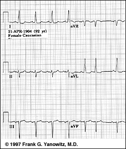
ECG with AF is above and below is an ECG with sinus rhythm

Remember that AF is a supraventricular tachyarrhythmia characterized by an ECG with absent P waves.

Causes for AF

Ischaemic heart disease
Thyrotoxicosis
Chronic rheumatic heart disease especially mitral stenosis

Post surgery
Chronic lung disease
Alcohol intoxication
Atrial septal defect

Types of Atrial Fibrillation

Remember that patients can have permanent AF ( it is unlikely the rhythm be reverted to sinus rhythm, you can anticipate this if patient has AF longer than 1 year, ECHO shows dilated left atrium ( >5cm)and patient is older. Patient may also have paroxysmal AF where there is possibility that the rhythm can be reverted to sinus rhythm.

Cardioversion

Cardioversion of AF to sinus rhythm can be done either electrically or pharmacologically,
However, remember that for elective cardioversion ( electrical or pharmacological), anticoagulate patients with AF >48 hours with warfarin 3-4 weeks before and after cardioversion.

Electrical cardioversion can be performed with synchronized DC shock of 50 till 100 J

Drugs used for Pharmacological Cardioversion

These drugs include amiodarone, flecanide, ibutilide, dofetilide, propafenone and quinidine. ( Learn more about amiodarone and flecanide)

Acute Management of Fast AF

Remember the principles below,

a) Sustain haemodynamic stability
b) Control ventricular rhythm
c) Anticoagulation
d) Find the underlying cause
e) Long term cardioversion ( rhythm control) or rate control

If a patient is admitted for fast atrial fibrillation, first thing to assess is patient’s haemodynamic stability, if patient’s blood pressure is unstable , urgent DC shock is needed. However, if the patient is stable and has fast AF, try to control the ventricular rhythm. Two common drugs are used, beta-blocker or digoxin, digoxin is preferred if patient has heart failure.
After the patient is stable, try to find the underlying cause for the problem. If you think patient has permanent AF or paroxysmal AF and planned for cardioversion, discuss anticoagulation ( warfarin) with the patient. You may just want to have rate control if you think patient is unlikely to be reverted back to sinus rhythm!

MRCP Questions

1)The following are recognised causes of atrial fibrillation EXCEPT


A anxiety
B hyperthyroidism
C atrial septal defect
D alcohol abuse
E COAD

ANSWER: A

2)A patient presenting with atrial fibrillation who has reverted to sinus rhythm is more likely to remain in sinus rhythm in the following circumstances:


A age >75 years old
B been commenced on warfarin
C left atrium size > 6 cm on ECHO
D short history on AF
E a ventricular rate on presentation of 130 bpm

ANSWER: D

3)Causes of atrial fibrillation include:


A Aortic stenosis
B Maladie de Roger VSD
C anxiety
D recent MI
E complete heart block

ANSWER: D

4) A 70 year-old lady presents with sudden breathlessness and chest discomfort. On examination, she was observed to have an irregular heart beat with rate of 140 bpm, BP 122/74 and normal heart sounds. On auscultation of the chest, Fine basal crepitations are heard. An ECG confirms fast. She is anticoagulated with heparin and given diuretics. Her heart rate remains rapid. What is the most appropriate management of the lady's AF?


1 ) Cardioversion.
2 ) IV amiodarone.
3 ) IV betablocker.
4 ) IV digoxin.
5 ) Oral quinidine therapy.

ANSWER: 4

5)A 70-year-old woman has a history of dyspnoea and palpitations for six months. An ECG at that time showed atrial fibrillation. She was given digoxin, diuretics and aspirin. She now presents with two short-lived episodes of altered sensation in the left face, left arm and leg. There is poor coordination of the left hand. ECHO was normal as was a CT head scan.

What is the most appropriate next step in management?


1 ) anticoagulation
2 ) carotid endarterectomy
3 ) clopidogrel
4 ) corticosteroid treatment
5 ) no action

ANSWER: 1


6)A 48 year-old MAN with atrial fibrillation is admitted for DC cardioversion.He is reverted to sinus rhythm. Which one of the following drugs would be most likely to maintain sinus rhythm following this procedure?

1 ) amiodarone
2 ) digoxin
3 ) heparin
4 ) sotalol
5 ) verapamil

ANSWER: 1

7)A 50-year-old politician presented with a strange fluttering sensation in his chest, but no chest pain. The symptoms had lasted 24 hours. An ECG revealed atrial fibrillation with a ventricular rate of 130 beats per minute.

Which one of the following drugs is most likely to restore sinus rhythm?

1 ) Adenosine
2 ) Bisoprolol
3 ) Digoxin
4 ) Flecainide
5 ) Verapamil

ANSWER: 4

8)The risk of embolic stroke with atrial fibrillation is increased in: EXCEPT

A Left ventricular ejection fraction of 60%.
B Diabetes mellitus.
C Controlled hypertension.
D Age greater than 75 years.
E Rheumatic mitral valve disease.

ANSWER: A

Tuesday, July 25, 2006

HEREDITARY HAEMOCHROMATOSIS

HEREDITARY HAEMOCHROMATOSIS (HH)

Today, I am going to talk about an important disease in gastroenterology. This disease is commonly asked in MRCP and popular questions being asked in MRCP Part 1 as well as Part 2 are as follow,

a) data interpretation for iron profile of a patient,
b) Presentations of hereditary haemochromatosis, usually a patient with non- specific joint pain,
c) Complications of HH,
d) HLA A3 association with the disease

Hereditary haemochhromatosis is a condition where there is abnormal high absorption of iron by the gut mucosa leading to iron overload in the body. It is another common cause of chronic liver disease in young age population besides Wilson’s disease. Remember that hereditary haemochromatosis is the commonest cause of iron overload in Western countries whereas in developing countries and Middle East, multiple frequent transfusions is the major cause of iron overload especially among patients with Thalassemia.

Data Interpretation For Hereditary Haemochromatosis

Features

Total iron increased
Ferritin increased
Fasting transferring saturation increased

Remember that due to iron overload in all organs especially endocrine organs such as pituitary, gonads, pancreas and also liver, cartilage, joints and etc this eventually leads to development of Diabetes mellitus, low testosterone and possibility of low libido and infertility.

Common presentations of Hereditary Haemochromatosis

Due to above reasons ( iron overload), patient may present with
- unspecific joint pain,
- chronic liver disease ( liver cirrhosis) and lead to liver cancer
- loss of libido and infertility
- DM

Complications of HH

Liver cirrhosis
Cardiomyopathy ( due to iron overload over cardiac muscle)

Treatment of HH

Recurrent phlebotomy, avoid Vit C ( can increase iron absorption) and alcohol.

Monday, July 24, 2006

RHEUMATIC FEVER

RHEUMATIC FEVER

Another popular topic in MRCP cardiology, common questions being asked in MRCP part 1 as well as part 2 are as follow,

a) Duckett Jones criteria to diagnose rheumatic fever,
b) Relationship between rheumatic fever and streptococcal infection,
c) Common presentations of rheumatic fever
d) Acute treatment for rheumatic fever

Why is rheumatic fever so important in MRCP? I think it is because the major cause of valvular heart diseases in developing countries is still chronic rheumatic disease.

DUCKETT JONES CRITERIA

The mnemonic to remember SPEC2 + WAFER2

Major Criteria

Subcutaneous nodule
Polyarthritis-described as migratory
Erythema marginatum
Carditis – pancarditis, usually in acute setting, no murmur can be heard, but there is possibility of mid-diastolic murmur ( Carey Coombes murmur)
Chorea- Sydenham’s chorea

Minor Criteria

White cells- raised ( leucocytosis)
Arthragia
Fever
ESR- raised or CRP
Rheumatic fever ( previous history)
PR interval prolonged

Diagnosis: Recent Streptococcal infection ( raised ASOT) + 2 majors or 1 major and 2 minors.

Streptococcal Infection and Rheumatic fever

Rheumatic fever usually occurs 3 weeks after Group A streptococcal infection.

Acute Treatment

Penicillin + aspirin ( high dose) and +/- steroid ( prednisolone)